1S84 | pdb_00001s84

PORCINE TRYPSIN COVALENT COMPLEX WITH 4-AMINO BUTANOL, BORATE AND ETHYLENE GLYCOL


Experimental Data Snapshot

  • Method: X-RAY DIFFRACTION
  • Resolution: 1.85 Å
  • R-Value Free: 
    0.177 (Depositor), 0.176 (DCC) 
  • R-Value Work: 
    0.155 (Depositor), 0.149 (DCC) 
  • R-Value Observed: 
    0.155 (Depositor) 

Starting Model: experimental
View more details

wwPDB Validation 3D Report Full Report

Validation slider image for 1S84

Ligand Structure Quality Assessment 


This is version 1.5 of the entry. See complete history

Literature

X-ray and NMR characterization of covalent complexes of trypsin, borate, and alcohols.

Transue, T.R.Krahn, J.M.Gabel, S.A.DeRose, E.F.London, R.E.

(2004) Biochemistry 43: 2829-2839

  • DOI: https://doi.org/10.1021/bi035782y
  • Primary Citation Related Structures: 
    1S5S, 1S6F, 1S6H, 1S81, 1S82, 1S83, 1S84, 1S85

  • PubMed Abstract: 

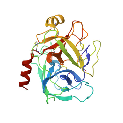
    An understanding of the physiological and toxicological properties of borate and the utilization of boronic acids in drug development require a basic understanding of borate-enzyme chemistry. We report here the extension of our recent NMR studies indicating the formation of a ternary borate-alcohol-trypsin complex. Crystallographic and solution state NMR studies of porcine trypsin were performed in the presence of borate and either of three alcohols designed to bind to the S1 affinity subsite: 4-aminobutanol, guanidine-3-propanol, and 4-hydroxymethylbenzamidine. Quaternary complexes of trypsin, borate, S1-binding alcohol, and ethylene glycol (a cryoprotectant), as well as a ternary trypsin, borate, and ethylene glycol complex have been observed in the crystalline state. Borate forms ester bonds to Ser195, ethylene glycol (two bonds), and the S1-binding alcohol (if present). Spectra from (1)H and (11)B NMR studies confirm that these complexes also exist in solution and also provide evidence for the formation of ternary trypsin, borate, and S1-subsite alcohol complexes which are not observed in the crystals using our experimental protocols. Analysis of eight crystal structures indicates that formation of an active site borate complex is in all cases accompanied by a significant (approximately 4%) increase in the b-axis dimension of the unit cell. Presumably, our inability to observe the ternary complexes in the crystalline state arises from the lower stability of these complexes and consequent inability to overcome the constraints imposed by the lattice contacts. A mechanism for the coupling of the lattice contacts with the active site that involves a conformational rearrangement of Gln192 is suggested. The structures presented here represent the first crystallographic demonstration of covalent binding of an enzyme by borate.


  • Organizational Affiliation
    • Laboratory of Structural Biology, National Institute of Environmental Health Sciences, National Institutes of Health, 111 T.W. Alexander Drive, Research Triangle Park, North Carolina 27709, USA.

Macromolecule Content 

  • Total Structure Weight: 24.35 kDa 
  • Atom Count: 2,047 
  • Modeled Residue Count: 223 
  • Deposited Residue Count: 223 
  • Unique protein chains: 1

Macromolecules

Find similar proteins by:|  3D Structure
Entity ID: 1
MoleculeChains  Sequence LengthOrganismDetailsImage
TRYPSIN223Sus scrofaMutation(s): 0 
EC: 3.4.21.4
UniProt
Find proteins for P00761 (Sus scrofa)
Explore P00761 
Go to UniProtKB:  P00761
Entity Groups
Sequence Clusters30% Identity50% Identity70% Identity90% Identity95% Identity100% Identity
UniProt GroupP00761
Sequence Annotations
Expand
Reference Sequence

Small Molecules

Ligands 4 Unique
IDChains Name / Formula / InChI Key2D Diagram3D Interactions
DYB

Query on DYB


Download:Ideal Coordinates CCD File
L [auth A]4-(1,3,2-DIOXABOROLAN-2-YLOXY)BUTAN-1-AMINIUM
C6 H15 B N O3
KTZDASPSWBAPAR-UHFFFAOYSA-O
SO4

Query on SO4


Download:Ideal Coordinates CCD File
C [auth A],
D [auth A],
E [auth A]
SULFATE ION
O4 S
QAOWNCQODCNURD-UHFFFAOYSA-L
EDO

Query on EDO


Download:Ideal Coordinates CCD File
F [auth A]
G [auth A]
H [auth A]
I [auth A]
J [auth A]
F [auth A],
G [auth A],
H [auth A],
I [auth A],
J [auth A],
K [auth A]
1,2-ETHANEDIOL
C2 H6 O2
LYCAIKOWRPUZTN-UHFFFAOYSA-N
CA

Query on CA


Download:Ideal Coordinates CCD File
B [auth A]CALCIUM ION
Ca
BHPQYMZQTOCNFJ-UHFFFAOYSA-N

Experimental Data & Validation

Experimental Data

  • Method: X-RAY DIFFRACTION
  • Resolution: 1.85 Å
  • R-Value Free:  0.177 (Depositor), 0.176 (DCC) 
  • R-Value Work:  0.155 (Depositor), 0.149 (DCC) 
  • R-Value Observed: 0.155 (Depositor) 
Space Group: P 21 21 21
Unit Cell:
Length ( Å )Angle ( ˚ )
a = 76.12α = 90
b = 55.771β = 90
c = 46.59γ = 90
Software Package:
Software NamePurpose
SCALEPACKdata scaling
CNSrefinement
CNSphasing

Structure Validation

View Full Validation Report


Ligand Structure Quality Assessment 


Entry History 

Deposition Data

Revision History  (Full details and data files)

  • Version 1.0: 2004-03-16
    Type: Initial release
  • Version 1.1: 2008-04-29
    Changes: Version format compliance
  • Version 1.2: 2011-07-13
    Changes: Version format compliance
  • Version 1.3: 2017-10-11
    Changes: Refinement description
  • Version 1.4: 2023-08-23
    Changes: Data collection, Database references, Derived calculations, Refinement description
  • Version 1.5: 2024-11-20
    Changes: Structure summary