1S56 | pdb_00001s56

Crystal Structure of "Truncated" Hemoglobin N (HbN) from Mycobacterium tuberculosis, Soaked with Xe Atoms


Experimental Data Snapshot

  • Method: X-RAY DIFFRACTION
  • Resolution: 2.43 Å
  • R-Value Free: 
    0.277 (Depositor), 0.284 (DCC) 
  • R-Value Work: 
    0.194 (Depositor), 0.202 (DCC) 
  • R-Value Observed: 
    0.198 (Depositor) 

Starting Model: experimental
View more details

wwPDB Validation 3D Report Full Report

Validation slider image for 1S56

Ligand Structure Quality Assessment 


This is version 1.3 of the entry. See complete history

Literature

Heme-ligand tunneling in group I truncated hemoglobins

Milani, M.Pesce, A.Ouellet, Y.Dewilde, S.Friedman, J.Ascenzi, P.Guertin, M.Bolognesi, M.

(2004) J Biological Chem 279: 21520-21525

  • DOI: https://doi.org/10.1074/jbc.M401320200
  • Primary Citation Related Structures: 
    1S56, 1S61, 1UVX, 1UVY

  • PubMed Abstract: 

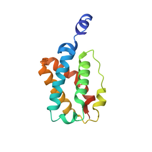
    Truncated hemoglobins (trHbs) are small hemoproteins forming a separate cluster within the hemoglobin superfamily; their functional roles in bacteria, plants, and unicellular eukaryotes are marginally understood. Crystallographic investigations have shown that the trHb fold (a two-on-two alpha-helical sandwich related to the globin fold) hosts a protein matrix tunnel system offering a potential path for ligand diffusion to the heme distal site. The tunnel topology is conserved in group I trHbs, although with modulation of its size/structure. Here, we present a crystallographic investigation on trHbs from Mycobacterium tuberculosis, Chlamydomonas eugametos, and Paramecium caudatum, showing that treatment of trHb crystals under xenon pressure leads to binding of xenon atoms at specific (conserved) sites along the protein matrix tunnel. The crystallographic results are in keeping with data from molecular dynamics simulations, where a dioxygen molecule is left free to diffuse within the protein matrix. Modulation of xenon binding over four main sites is related to the structural properties of the tunnel system in the three trHbs and may be connected to their functional roles. In a parallel crystallographic investigation on M. tuberculosis trHbN, we show that butyl isocyanide also binds within the apolar tunnel, in excellent agreement with concepts derived from the xenon binding experiments. These results, together with recent data on atypical CO rebinding kinetics to group I trHbs, underline the potential role of the tunnel system in supporting diffusion, but also accumulation in multiple copies, of low polarity ligands/molecules within group I trHbs.


  • Organizational Affiliation
    • Giannina Gaslini Institute, Largo G. Gaslini, 5. 16147 Genoa, Italy.

Macromolecule Content 

  • Total Structure Weight: 31.4 kDa 
  • Atom Count: 2,179 
  • Modeled Residue Count: 263 
  • Deposited Residue Count: 272 
  • Unique protein chains: 1

Macromolecules

Find similar proteins by:|  3D Structure
Entity ID: 1
MoleculeChains  Sequence LengthOrganismDetailsImage
Hemoglobin-like protein HbN
A, B
136Mycobacterium tuberculosisMutation(s): 0 
Gene Names: GLBNRV1542CMT1594MTCY48.23MB1569C
UniProt
Find proteins for P9WN25 (Mycobacterium tuberculosis (strain ATCC 25618 / H37Rv))
Explore P9WN25 
Go to UniProtKB:  P9WN25
Entity Groups
Sequence Clusters30% Identity50% Identity70% Identity90% Identity95% Identity100% Identity
UniProt GroupP9WN25
Sequence Annotations
Expand
Reference Sequence

Small Molecules

Ligands 6 Unique
IDChains Name / Formula / InChI Key2D Diagram3D Interactions
HEC

Query on HEC


Download:Ideal Coordinates CCD File
E [auth A]HEME C
C34 H34 Fe N4 O4
HXQIYSLZKNYNMH-LJNAALQVSA-N
HEM

Query on HEM


Download:Ideal Coordinates CCD File
L [auth B]PROTOPORPHYRIN IX CONTAINING FE
C34 H32 Fe N4 O4
KABFMIBPWCXCRK-RGGAHWMASA-L
XE

Query on XE


Download:Ideal Coordinates CCD File
F [auth A]
G [auth A]
H [auth A]
I [auth A]
M [auth B]
F [auth A],
G [auth A],
H [auth A],
I [auth A],
M [auth B],
N [auth B],
O [auth B],
P [auth B]
XENON
Xe
FHNFHKCVQCLJFQ-UHFFFAOYSA-N
PO4

Query on PO4


Download:Ideal Coordinates CCD File
K [auth B]PHOSPHATE ION
O4 P
NBIIXXVUZAFLBC-UHFFFAOYSA-K
K

Query on K


Download:Ideal Coordinates CCD File
D [auth A]POTASSIUM ION
K
NPYPAHLBTDXSSS-UHFFFAOYSA-N
CYN

Query on CYN


Download:Ideal Coordinates CCD File
C [auth A],
J [auth B]
CYANIDE ION
C N
XFXPMWWXUTWYJX-UHFFFAOYSA-N

Experimental Data & Validation

Experimental Data

  • Method: X-RAY DIFFRACTION
  • Resolution: 2.43 Å
  • R-Value Free:  0.277 (Depositor), 0.284 (DCC) 
  • R-Value Work:  0.194 (Depositor), 0.202 (DCC) 
  • R-Value Observed: 0.198 (Depositor) 
Space Group: P 21 21 21
Unit Cell:
Length ( Å )Angle ( ˚ )
a = 44.321α = 90
b = 61.959β = 90
c = 90.747γ = 90
Software Package:
Software NamePurpose
REFMACrefinement
MOSFLMdata reduction
CCP4data scaling
EPMRphasing

Structure Validation

View Full Validation Report


Ligand Structure Quality Assessment 


Entry History 

Deposition Data

Revision History  (Full details and data files)

  • Version 1.0: 2004-06-29
    Type: Initial release
  • Version 1.1: 2008-04-29
    Changes: Version format compliance
  • Version 1.2: 2011-07-13
    Changes: Version format compliance
  • Version 1.3: 2023-08-23
    Changes: Data collection, Database references, Derived calculations, Refinement description