1S3T | pdb_00001s3t

BORATE INHIBITED BACILLUS PASTEURII UREASE CRYSTAL STRUCTURE


Experimental Data Snapshot

  • Method: X-RAY DIFFRACTION
  • Resolution: 2.10 Å
  • R-Value Free: 
    0.205 (Depositor), 0.208 (DCC) 
  • R-Value Work: 
    0.177 (Depositor), 0.188 (DCC) 
  • R-Value Observed: 
    0.178 (Depositor) 

Starting Model: experimental
View more details

wwPDB Validation 3D Report Full Report

Validation slider image for 1S3T

This is version 1.5 of the entry. See complete history

Literature

Molecular Details of Urease Inhibition by Boric Acid: Insights into the Catalytic Mechanism.

Benini, S.Rypniewski, W.R.Wilson, K.S.Mangani, S.Ciurli, S.

(2004) J Am Chem Soc 126: 3714-3715

  • DOI: https://doi.org/10.1021/ja049618p
  • Primary Citation Related Structures: 
    1S3T

  • PubMed Abstract: 

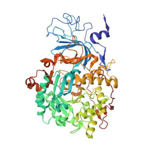
    The structure of the complex of urease, a Ni-containing metalloenzyme, with boric acid was determined at 2.10 A resolution. The complex shows the unprecedented binding mode of the competitive inhibitor to the dinuclear metal center, with the B(OH)3 molecule bridging the Ni ions and leaving in place the bridging hydroxide. Boric acid can be considered a substrate analogue of urea, and the structure supports the proposal that the Ni-bridging hydroxide acts as the nucleophile in the enzymatic process of urea hydrolysis.


  • Organizational Affiliation
    • York Structural Biology Laboratory, University of York, York, UK.

Macromolecule Content 

  • Total Structure Weight: 87.1 kDa 
  • Atom Count: 6,376 
  • Modeled Residue Count: 792 
  • Deposited Residue Count: 796 
  • Unique protein chains: 3

Macromolecules

Find similar proteins by:|  3D Structure
Entity ID: 1
MoleculeChains  Sequence LengthOrganismDetailsImage
Urease gamma subunit100Sporosarcina pasteuriiMutation(s): 1 
EC: 3.5.1.5
UniProt
Find proteins for P41022 (Sporosarcina pasteurii)
Explore P41022 
Go to UniProtKB:  P41022
Entity Groups
Sequence Clusters30% Identity50% Identity70% Identity90% Identity95% Identity100% Identity
UniProt GroupP41022
Sequence Annotations
Expand
Reference Sequence
Find similar proteins by:|  3D Structure
Entity ID: 2
MoleculeChains  Sequence LengthOrganismDetailsImage
Urease beta subunit126Sporosarcina pasteuriiMutation(s): 0 
EC: 3.5.1.5
UniProt
Find proteins for P41021 (Sporosarcina pasteurii)
Explore P41021 
Go to UniProtKB:  P41021
Entity Groups
Sequence Clusters30% Identity50% Identity70% Identity90% Identity95% Identity100% Identity
UniProt GroupP41021
Sequence Annotations
Expand
Reference Sequence
Find similar proteins by:|  3D Structure
Entity ID: 3
MoleculeChains  Sequence LengthOrganismDetailsImage
Urease alpha subunit570Sporosarcina pasteuriiMutation(s): 1 
EC: 3.5.1.5
UniProt
Find proteins for P41020 (Sporosarcina pasteurii)
Explore P41020 
Go to UniProtKB:  P41020
Entity Groups
Sequence Clusters30% Identity50% Identity70% Identity90% Identity95% Identity100% Identity
UniProt GroupP41020
Sequence Annotations
Expand
Reference Sequence

Experimental Data & Validation

Experimental Data

  • Method: X-RAY DIFFRACTION
  • Resolution: 2.10 Å
  • R-Value Free:  0.205 (Depositor), 0.208 (DCC) 
  • R-Value Work:  0.177 (Depositor), 0.188 (DCC) 
  • R-Value Observed: 0.178 (Depositor) 
Space Group: P 63 2 2
Unit Cell:
Length ( Å )Angle ( ˚ )
a = 130.906α = 90
b = 130.906β = 90
c = 189.366γ = 120
Software Package:
Software NamePurpose
REFMACrefinement
DENZOdata reduction
SCALEPACKdata scaling
AMoREphasing

Structure Validation

View Full Validation Report


Entry History 

Deposition Data

Revision History  (Full details and data files)

  • Version 1.0: 2004-04-06
    Type: Initial release
  • Version 1.1: 2008-04-29
    Changes: Version format compliance
  • Version 1.2: 2011-07-13
    Changes: Derived calculations, Version format compliance
  • Version 1.3: 2015-07-08
    Changes: Source and taxonomy
  • Version 1.4: 2023-08-23
    Changes: Data collection, Database references, Derived calculations, Refinement description
  • Version 1.5: 2023-11-15
    Changes: Data collection