1S1Y | pdb_00001s1y

Photoactivated chromophore conformation in Photoactive Yellow Protein (E46Q mutant) from 10 microseconds to 3 milliseconds


Experimental Data Snapshot

  • Method: X-RAY DIFFRACTION
  • Resolution: 1.60 Å
  • R-Value Free: 
    0.070 (Depositor) 
  • R-Value Work: 
    0.064 (Depositor) 
  • R-Value Observed: 
    0.066 (Depositor) 

wwPDB Validation 3D Report Full Report

Validation slider image for 1S1Y

This is version 1.4 of the entry. See complete history

Literature

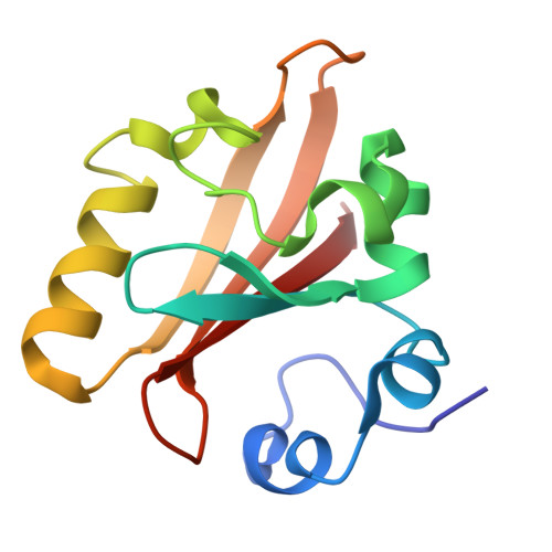
Chromophore conformation and the evolution of tertiary structural changes in photoactive yellow protein

Anderson, S.Srajer, V.Pahl, R.Rajagopal, S.Schotte, F.Anfinrud, P.Wulff, M.Moffat, K.

(2004) Structure 12: 1039-1045

  • DOI: https://doi.org/10.1016/j.str.2004.04.008
  • Primary Citation Related Structures: 
    1S1Y, 1S1Z

  • PubMed Abstract: 

    We use time-resolved crystallography to observe the structural progression of a bacterial blue light photoreceptor throughout its photocycle. Data were collected from 10 ns to 100 ms after photoactivation of the E46Q mutant of photoactive yellow protein. Refinement of transient chromophore conformations shows that the spectroscopically distinct intermediates are formed via progressive disruption of the hydrogen bond network to the chromophore. Although structural change occurs within a few nanoseconds on and around the chromophore, it takes milliseconds for a distinct pattern of tertiary structural change to fully progress through the entire molecule, thus generating the putative signaling state. Remarkably, the coupling between the chromophore conformation and the tertiary structure of this small protein is not tight: there are leads and lags between changes in the conformation of the chromophore and the protein tertiary structure.


  • Organizational Affiliation
    • Department of Biochemistry and Molecular Biology, University of Chicago, Chicago, IL 60637, USA. smander@midway.uchicago.edu

Macromolecule Content 

  • Total Structure Weight: 14.05 kDa 
  • Atom Count: 1,355 
  • Modeled Residue Count: 125 
  • Deposited Residue Count: 125 
  • Unique protein chains: 1

Macromolecules

Find similar proteins by:|  3D Structure
Entity ID: 1
MoleculeChains  Sequence LengthOrganismDetailsImage
Photoactive yellow protein125Halorhodospira halophilaMutation(s): 1 
Gene Names: PYP
UniProt
Find proteins for P16113 (Halorhodospira halophila)
Explore P16113 
Go to UniProtKB:  P16113
Entity Groups
Sequence Clusters30% Identity50% Identity70% Identity90% Identity95% Identity100% Identity
UniProt GroupP16113
Sequence Annotations
Expand
Reference Sequence

Small Molecules

Ligands 1 Unique
IDChains Name / Formula / InChI Key2D Diagram3D Interactions
HC4

Query on HC4


Download:Ideal Coordinates CCD File
B [auth A]4'-HYDROXYCINNAMIC ACID
C9 H8 O3
NGSWKAQJJWESNS-ZZXKWVIFSA-N

Experimental Data & Validation

Experimental Data

  • Method: X-RAY DIFFRACTION
  • Resolution: 1.60 Å
  • R-Value Free:  0.070 (Depositor) 
  • R-Value Work:  0.064 (Depositor) 
  • R-Value Observed: 0.066 (Depositor) 
Space Group: P 63
Unit Cell:
Length ( Å )Angle ( ˚ )
a = 66.917α = 90
b = 66.917β = 90
c = 41.002γ = 120
Software Package:
Software NamePurpose
LaueViewdata scaling
LaueViewdata reduction
XTALVIEWrefinement
SHELXL-97refinement

Structure Validation

View Full Validation Report


Entry History 

Deposition Data

Revision History  (Full details and data files)

  • Version 1.0: 2004-06-15
    Type: Initial release
  • Version 1.1: 2008-04-29
    Changes: Version format compliance
  • Version 1.2: 2011-07-13
    Changes: Version format compliance
  • Version 1.3: 2017-10-11
    Changes: Refinement description
  • Version 1.4: 2021-10-27
    Changes: Database references, Derived calculations