1S1O | pdb_00001s1o

NMR Structure of a D,L Alternating pentadecamer of norleucine: double antiparallel beta-helix


Experimental Data Snapshot

  • Method: SOLUTION NMR
  • Conformers Calculated: 50 
  • Conformers Submitted: 
  • Selection Criteria: structures with the lowest energy 

wwPDB Validation 3D Report Full Report

Validation slider image for 1S1O

This is version 2.0 of the entry. See complete history

Literature

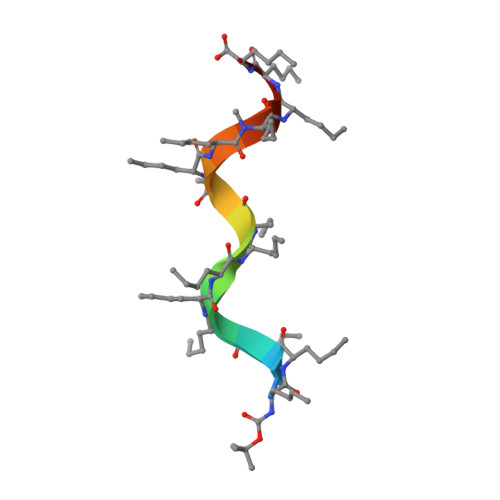
Solution structure of a D,L-alternating oligonorleucine as a model of double-stranded antiparallel beta-helix.

Navarro, E.Fenude, E.Celda, B.

(2002) Biopolymers 64: 198-209

  • DOI: https://doi.org/10.1002/bip.10172
  • Primary Citation Related Structures: 
    1S1O

  • PubMed Abstract: 

    Conformational characteristics of alternating D,L linear peptides are of particular interest because of their capacity to form transmembrane channels with different transport properties, as some natural antibiotics do. Single- and double-stranded beta-helical structures are common for alternating D,L peptides. The stability of the beta-helix depends on several structural factors, such as the backbone peptide length, type and position of side chains, and nature of terminal groups. The NMR and molecular dynamics solution conformation of a synthetic alternating D,L-oligopeptide with 15 norleucines (XVMe) has been used as a model to get insight in to the conformational features of double-stranded beta-helix structures. The NH chemical shift values (delta(NH)) and long-range nuclear Overhauser effects (NOE) cross peaks, in particular interstrand connectivities, clearly point to an antiparallel double-stranded beta-helix for the XVMe major conformation in solution. An extensive set of distances (from NOE cross peaks) and H-bonds (from delta(NH)) has been included in the molecular dynamics calculations. The experimental NMR data and theoretical calculations clearly indicate that the most probable conformation of XVMe in solution is a double-strand antiparallel beta(5.6) increasing decreasing-helix structure.


  • Organizational Affiliation
    • Departamento de Química Física, Facultat de Químicas, Universitat de Valencia, C/Dr. Moliner,50, 46100-Burjassot (Valencia), Spain.

Macromolecule Content 

  • Total Structure Weight: 3.69 kDa 
  • Atom Count: 258 
  • Modeled Residue Count: 32 
  • Deposited Residue Count: 32 
  • Unique protein chains: 1

Macromolecules

Find similar proteins by:  Sequence   |   3D Structure  
Entity ID: 1
MoleculeChains  Sequence LengthOrganismDetailsImage
BOC-L-NLE-(D-NLE-L-NLE)5-D-NLE(METHYL)-L-NLE-D-NLE-L-NLE METHYL ESTER
A, B
16N/AMutation(s): 0 

Small Molecules

Modified Residues  2 Unique
IDChains TypeFormula2D DiagramParent
DNE
Query on DNE
A, B
D-PEPTIDE LINKINGC6 H13 N O2

--

DNM
Query on DNM
A, B
D-PEPTIDE LINKINGC7 H15 N O2

--

NLE
Query on NLE
A, B
L-PEPTIDE LINKINGC6 H13 N O2LEU
NLO
Query on NLO
A, B
L-PEPTIDE LINKINGC7 H15 N O2LEU

Biologically Interesting Molecules (External Reference) 

1 Unique

Experimental Data & Validation

Experimental Data

  • Method: SOLUTION NMR
  • Conformers Calculated: 50 
  • Conformers Submitted: 
  • Selection Criteria: structures with the lowest energy 

Structure Validation

View Full Validation Report


Entry History 

Deposition Data

Revision History  (Full details and data files)

  • Version 1.0: 2004-02-24
    Type: Initial release
  • Version 1.1: 2008-04-29
    Changes: Version format compliance
  • Version 1.2: 2011-07-13
    Changes: Version format compliance
  • Version 1.3: 2012-09-19
    Changes: Non-polymer description, Structure summary
  • Version 1.4: 2012-12-12
    Changes: Other
  • Version 1.5: 2022-02-09
    Changes: Data collection, Database references, Derived calculations
  • Version 2.0: 2023-11-15
    Changes: Atomic model, Data collection, Derived calculations