1S0P | pdb_00001s0p

Structure of the N-Terminal Domain of the Adenylyl Cyclase-Associated Protein (CAP) from Dictyostelium discoideum.


Experimental Data Snapshot

  • Method: X-RAY DIFFRACTION
  • Resolution: 1.40 Å
  • R-Value Free: 
    0.210 (Depositor) 
  • R-Value Work: 
    0.180 (Depositor) 
  • R-Value Observed: 
    0.260 (Depositor) 

wwPDB Validation 3D Report Full Report

Validation slider image for 1S0P

This is version 1.3 of the entry. See complete history

Literature

STRUCTURE OF THE N-TERMINAL DOMAIN OF THE ADENYLYL CYCLASE-ASSOCIATED PROTEIN (CAP) FROM DICTYOSTELIUM DISCOIDEUM

Ksiazek, D.Brandstetter, H.Israel, L.Bourenkov, G.P.Katchalova, G.Janssen, K.P.Bartunik, H.D.Noegel, A.A.Schleicher, M.Holak, T.A.

(2003) Structure 11: 1171-1178

  • DOI: https://doi.org/10.1016/s0969-2126(03)00180-1
  • Primary Citation Related Structures: 
    1S0P

  • PubMed Abstract: 

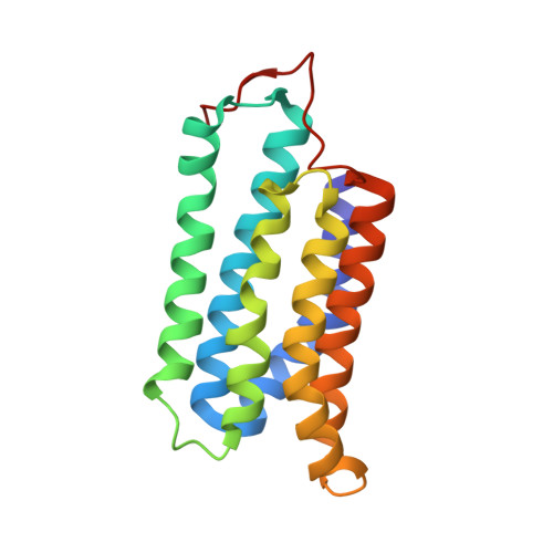
    Cyclase-associated proteins (CAPs) are widely distributed and highly conserved proteins that regulate actin remodeling in response to cellular signals. The N termini of CAPs play a role in Ras signaling and bind adenylyl cyclase; the C termini bind to G-actin and thereby alter the dynamic rearrangements of the microfilament system. We report here the X-ray structure of the core of the N-terminal domain of the CAP from Dictyostelium discoideum, which comprises residues 51-226, determined by a combination of single isomorphous replacement with anomalous scattering (SIRAS). The overall structure of this fragment is an alpha helix bundle composed of six antiparallel helices. Results from gel filtration and crosslinking experiments for CAP(1-226), CAP(255-464), and the full-length protein, together with the CAP N-terminal domain structure and the recently determined CAP C-terminal domain structure, provide evidence that the functional structure of CAP is multimeric.


  • Organizational Affiliation
    • Max-Planck Institut für Biochemie, 82152 Martinsried, Germany.

Macromolecule Content 

  • Total Structure Weight: 39.38 kDa 
  • Atom Count: 3,379 
  • Modeled Residue Count: 352 
  • Deposited Residue Count: 352 
  • Unique protein chains: 1

Macromolecules

Find similar proteins by:|  3D Structure
Entity ID: 1
MoleculeChains  Sequence LengthOrganismDetailsImage
Adenylyl cyclase-associated protein
A, B
176Dictyostelium discoideumMutation(s): 0 
Gene Names: CAP
UniProt
Find proteins for P54654 (Dictyostelium discoideum)
Explore P54654 
Go to UniProtKB:  P54654
Entity Groups
Sequence Clusters30% Identity50% Identity70% Identity90% Identity95% Identity100% Identity
UniProt GroupP54654
Sequence Annotations
Expand
Reference Sequence

Small Molecules

Ligands 1 Unique
IDChains Name / Formula / InChI Key2D Diagram3D Interactions
MG

Query on MG


Download:Ideal Coordinates CCD File
C [auth A]MAGNESIUM ION
Mg
JLVVSXFLKOJNIY-UHFFFAOYSA-N

Experimental Data & Validation

Experimental Data

  • Method: X-RAY DIFFRACTION
  • Resolution: 1.40 Å
  • R-Value Free:  0.210 (Depositor) 
  • R-Value Work:  0.180 (Depositor) 
  • R-Value Observed: 0.260 (Depositor) 
Space Group: P 1
Unit Cell:
Length ( Å )Angle ( ˚ )
a = 37.52α = 97.42
b = 42.13β = 105.09
c = 53.74γ = 97.15
Software Package:
Software NamePurpose
DENZOdata reduction
SCALEPACKdata scaling
CNSrefinement
CNSphasing

Structure Validation

View Full Validation Report


Entry History 

Deposition Data

Revision History  (Full details and data files)

  • Version 1.0: 2004-01-27
    Type: Initial release
  • Version 1.1: 2008-04-29
    Changes: Version format compliance
  • Version 1.2: 2011-07-13
    Changes: Version format compliance
  • Version 1.3: 2024-02-14
    Changes: Data collection, Database references, Derived calculations