1RWY | pdb_00001rwy

CRYSTAL STRUCTURE OF RAT ALPHA-PARVALBUMIN AT 1.05 RESOLUTION


Experimental Data Snapshot

  • Method: X-RAY DIFFRACTION
  • Resolution: 1.05 Å
  • R-Value Free: 
    0.162 (Depositor), 0.167 (DCC) 
  • R-Value Work: 
    0.140 (DCC) 
  • R-Value Observed: 
    0.133 (Depositor) 

Starting Model: experimental
View more details

wwPDB Validation 3D Report Full Report

Validation slider image for 1RWY

This is version 1.5 of the entry. See complete history

Literature

Crystal Structure of Rat Alpha-Parvalbumin at 1.05 Resolution

Bottoms, C.A.Schuermann, J.P.Agah, S.Henzl, M.T.Tanner, J.J.

(2004) Protein Sci 13: 1724-1734

  • DOI: https://doi.org/10.1110/ps.03571004
  • Primary Citation Related Structures: 
    1RWY

  • PubMed Abstract: 

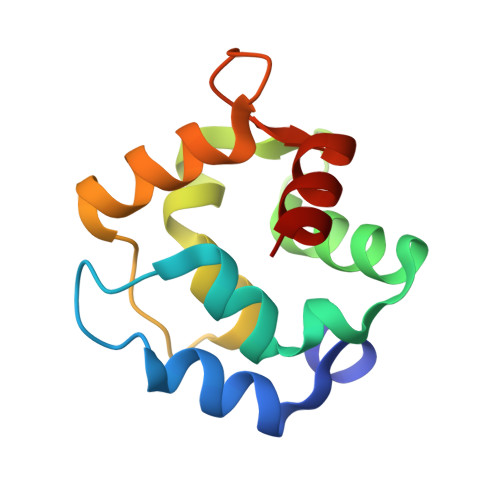
    The crystal structure of rat alpha-parvalbumin has been determined at 1.05 Angstrom resolution, using synchrotron data collected at Advanced Photon Source beamline 19-ID. After refinement with SHELX, employing anisotropic displacement parameters and riding hydrogen atoms, R = 0.132 and R(free) = 0.162. The average coordinate estimated standard deviations are 0.021 Angstrom and 0.038 Angstrom for backbone atoms and side-chain atoms, respectively. Besides providing a more precise view of the alpha-isoform than previously available, these data permit comparison with the 0.91 Angstrom structure determined for pike beta-parvalbumin. Visualization of the anisotropic displacement parameters as thermal ellipsoids yields insight into the atomic motion within the Ca(2+)-binding sites. The asymmetric unit includes three parvalbumin (PV) molecules. Interestingly, the EF site in one displays uncharacteristic flexibility. The ellipsoids for Asp-92 are particularly large and non-spherical, and the shape of the Ca(2+) ellipsoid implies significant vibrational motion perpendicular to the plane defined by the four y and z ligands. The relative dearth of crystal-packing interactions in this site suggests that the heightened flexibility may be the result of diminished intermolecular contacts. The implication is that, by impeding conformational mobility, crystal-packing forces may cause serious overestimation of EF-hand rigidity. The high quality of the data permitted 11 residues to be modeled in alternative side-chain conformations, including the two core residues, Ile-97 and Leu-105. The discrete disorder observed for Ile-97 may have functional ramifications, providing a mechanism for communicating binding status between the CD and EF binding loops and between the PV metal ion-binding domain and the N-terminal AB region.


  • Organizational Affiliation
    • Department of Chemistry, University of Missouri-Columbia, Columbia, MO 65211, USA.

Macromolecule Content 

  • Total Structure Weight: 36.34 kDa 
  • Atom Count: 2,932 
  • Modeled Residue Count: 327 
  • Deposited Residue Count: 327 
  • Unique protein chains: 1

Macromolecules

Find similar proteins by:|  3D Structure
Entity ID: 1
MoleculeChains  Sequence LengthOrganismDetailsImage
PARVALBUMIN ALPHA
A, B, C
109Rattus norvegicusMutation(s): 0 
Gene Names: PVALBPVA
UniProt
Find proteins for P02625 (Rattus norvegicus)
Explore P02625 
Go to UniProtKB:  P02625
Entity Groups
Sequence Clusters30% Identity50% Identity70% Identity90% Identity95% Identity100% Identity
UniProt GroupP02625
Sequence Annotations
Expand
Reference Sequence

Small Molecules

Ligands 5 Unique
IDChains Name / Formula / InChI Key2D Diagram3D Interactions
PG4

Query on PG4


Download:Ideal Coordinates CCD File
P [auth C]TETRAETHYLENE GLYCOL
C8 H18 O5
UWHCKJMYHZGTIT-UHFFFAOYSA-N
SO4

Query on SO4


Download:Ideal Coordinates CCD File
F [auth A],
J [auth B],
K [auth B],
L [auth B]
SULFATE ION
O4 S
QAOWNCQODCNURD-UHFFFAOYSA-L
ACY

Query on ACY


Download:Ideal Coordinates CCD File
G [auth A]ACETIC ACID
C2 H4 O2
QTBSBXVTEAMEQO-UHFFFAOYSA-N
CA

Query on CA


Download:Ideal Coordinates CCD File
D [auth A]
E [auth A]
H [auth B]
I [auth B]
M [auth C]
D [auth A],
E [auth A],
H [auth B],
I [auth B],
M [auth C],
N [auth C]
CALCIUM ION
Ca
BHPQYMZQTOCNFJ-UHFFFAOYSA-N
NH4

Query on NH4


Download:Ideal Coordinates CCD File
O [auth C]AMMONIUM ION
H4 N
QGZKDVFQNNGYKY-UHFFFAOYSA-O

Experimental Data & Validation

Experimental Data

  • Method: X-RAY DIFFRACTION
  • Resolution: 1.05 Å
  • R-Value Free:  0.162 (Depositor), 0.167 (DCC) 
  • R-Value Work:  0.140 (DCC) 
  • R-Value Observed: 0.133 (Depositor) 
Space Group: P 21 21 21
Unit Cell:
Length ( Å )Angle ( ˚ )
a = 33.838α = 90
b = 54.708β = 90
c = 153.643γ = 90
Software Package:
Software NamePurpose
SHELXL-97refinement
DENZOdata reduction
HKL-2000data reduction
SCALEPACKdata scaling
CNSrefinement
SHELXmodel building
HKL-2000data collection
HKL-2000data scaling
CNSphasing
SHELXphasing

Structure Validation

View Full Validation Report


Entry History 

Deposition Data

Revision History  (Full details and data files)

  • Version 1.0: 2004-05-11
    Type: Initial release
  • Version 1.1: 2007-10-16
    Changes: Version format compliance
  • Version 1.2: 2011-07-13
    Changes: Version format compliance
  • Version 1.3: 2017-10-11
    Changes: Advisory, Refinement description
  • Version 1.4: 2019-07-24
    Changes: Data collection, Refinement description
  • Version 1.5: 2023-08-23
    Changes: Advisory, Data collection, Database references, Derived calculations, Refinement description