1RWI | pdb_00001rwi

Extracellular domain of Mycobacterium tuberculosis PknD


Experimental Data Snapshot

  • Method: X-RAY DIFFRACTION
  • Resolution: 1.80 Å
  • R-Value Free: 
    0.220 (Depositor) 
  • R-Value Work: 
    0.190 (Depositor) 
  • R-Value Observed: 
    0.193 (Depositor) 

wwPDB Validation 3D Report Full Report

Validation slider image for 1RWI

This is version 1.3 of the entry. See complete history

Literature

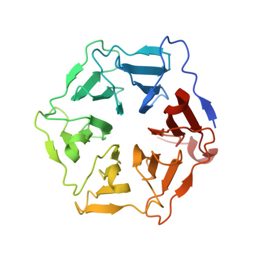
Sensor Domain of the Mycobacterium tuberculosis Receptor Ser/Thr Protein Kinase, PknD, forms a Highly Symmetric beta Propeller.

Good, M.C.Greenstein, A.E.Young, T.A.Ng, H.L.Alber, T.

(2004) J Mol Biology 339: 459-469

  • DOI: https://doi.org/10.1016/j.jmb.2004.03.063
  • Primary Citation Related Structures: 
    1RWI, 1RWL

  • PubMed Abstract: 

    Diverse pathogenic bacteria produce transmembrane receptor Ser/Thr protein kinases (STPKs), but little is known about the signals mediated by these "eukaryotic-like" proteins. To explore the basis for signaling in the bacterial STPK receptor family, we determined the structure of the sensor domain of Mycobacterium tuberculosis PknD. In two crystal forms, the PknD sensor domain forms a rigid, six-bladed beta-propeller with a flexible tether to the transmembrane domain. The PknD sensor domain is the most symmetric beta-propeller structure described. All residues that vary most among the blade subdomains cluster in the large "cup" motif, analogous to the ligand-binding surface in many beta-propeller proteins. These results suggest that PknD binds a multivalent ligand that signals by changing the quaternary structure of the intracellular kinase domain.


  • Organizational Affiliation
    • Department of Molecular and Cell Biology, University of California, 339 Hildebrand Hall #3206, Berkeley, CA 94720-3206, USA.

Macromolecule Content 

  • Total Structure Weight: 58.61 kDa 
  • Atom Count: 3,992 
  • Modeled Residue Count: 510 
  • Deposited Residue Count: 540 
  • Unique protein chains: 1

Macromolecules

Find similar proteins by:|  3D Structure
Entity ID: 1
MoleculeChains  Sequence LengthOrganismDetailsImage
Serine/threonine-protein kinase pknDA [auth B],
B [auth A]
270Mycobacterium tuberculosisMutation(s): 0 
Gene Names: PKNDRV0931CMT0958MTCY08C9.08
EC: 2.7.1.37 (PDB Primary Data), 2.7.11.1 (UniProt)
UniProt
Find proteins for P9WI79 (Mycobacterium tuberculosis (strain ATCC 25618 / H37Rv))
Explore P9WI79 
Go to UniProtKB:  P9WI79
Entity Groups
Sequence Clusters30% Identity50% Identity70% Identity90% Identity95% Identity100% Identity
UniProt GroupP9WI79
Sequence Annotations
Expand
Reference Sequence

Small Molecules

Ligands 1 Unique
IDChains Name / Formula / InChI Key2D Diagram3D Interactions
CD

Query on CD


Download:Ideal Coordinates CCD File
C [auth B]
D [auth B]
E [auth B]
F [auth B]
G [auth B]
C [auth B],
D [auth B],
E [auth B],
F [auth B],
G [auth B],
H [auth B],
I [auth B],
J [auth B],
K [auth B],
L [auth B],
M [auth A],
N [auth A],
O [auth A],
P [auth A]
CADMIUM ION
Cd
WLZRMCYVCSSEQC-UHFFFAOYSA-N

Experimental Data & Validation

Experimental Data

  • Method: X-RAY DIFFRACTION
  • Resolution: 1.80 Å
  • R-Value Free:  0.220 (Depositor) 
  • R-Value Work:  0.190 (Depositor) 
  • R-Value Observed: 0.193 (Depositor) 
Space Group: P 1 21 1
Unit Cell:
Length ( Å )Angle ( ˚ )
a = 43.94α = 90
b = 94.58β = 98.1
c = 71.77γ = 90
Software Package:
Software NamePurpose
MOSFLMdata reduction
SCALAdata scaling
DMmodel building
REFMACrefinement
CCP4data scaling
DMphasing

Structure Validation

View Full Validation Report


Entry History 

Deposition Data

Revision History  (Full details and data files)

  • Version 1.0: 2004-04-27
    Type: Initial release
  • Version 1.1: 2008-04-29
    Changes: Version format compliance
  • Version 1.2: 2011-07-13
    Changes: Version format compliance
  • Version 1.3: 2024-02-14
    Changes: Data collection, Database references, Derived calculations