1RPL | pdb_00001rpl

2.3 ANGSTROMS CRYSTAL STRUCTURE OF THE CATALYTIC DOMAIN OF DNA POLYMERASE BETA


Experimental Data Snapshot

  • Method: X-RAY DIFFRACTION
  • Resolution: 2.30 Å
  • R-Value Work: 
    0.220 (Depositor) 
  • R-Value Observed: 
    0.220 (Depositor) 

wwPDB Validation 3D Report Full Report

Validation slider image for 1RPL

This is version 1.4 of the entry. See complete history

Literature

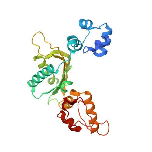
2.3 A crystal structure of the catalytic domain of DNA polymerase beta.

Davies 2nd., J.F.Almassy, R.J.Hostomska, Z.Ferre, R.A.Hostomsky, Z.

(1994) Cell 76: 1123-1133

  • DOI: https://doi.org/10.1016/0092-8674(94)90388-3
  • Primary Citation Related Structures: 
    1RPL

  • PubMed Abstract: 

    The crystal structure of the catalytic domain of rat DNA polymerase beta (pol beta) has been determined at 2.3 A resolution and refined to an R factor of 0.22. The mixed alpha/beta protein has three subdomains arranged in an overall U shape reminiscent of other polymerase structures. The folding topology of pol beta, however, is unique. Two divalent metals bind near three aspartic acid residues implicated in the catalytic activity. In the presence of Mn2+ and dTTP, interpretable electron density is seen for two metals and the triphosphate, but not the deoxythymidine moiety. The principal interaction of the triphosphate moiety is with the bound divalent metals.


  • Organizational Affiliation
    • Agouron Pharmaceuticals, Incorporated, San Diego, California 92121.

Macromolecule Content 

  • Total Structure Weight: 29.15 kDa 
  • Atom Count: 2,024 
  • Modeled Residue Count: 241 
  • Deposited Residue Count: 251 
  • Unique protein chains: 1

Macromolecules

Find similar proteins by:|  3D Structure
Entity ID: 1
MoleculeChains  Sequence LengthOrganismDetailsImage
DNA POLYMERASE BETA251Rattus norvegicusMutation(s): 0 
EC: 2.7.7.7 (UniProt), 4.2.99.18 (UniProt), 4.2.99 (UniProt)
UniProt
Find proteins for P06766 (Rattus norvegicus)
Explore P06766 
Go to UniProtKB:  P06766
Entity Groups
Sequence Clusters30% Identity50% Identity70% Identity90% Identity95% Identity100% Identity
UniProt GroupP06766
Sequence Annotations
Expand
Reference Sequence

Experimental Data & Validation

Experimental Data

  • Method: X-RAY DIFFRACTION
  • Resolution: 2.30 Å
  • R-Value Work:  0.220 (Depositor) 
  • R-Value Observed: 0.220 (Depositor) 
Space Group: P 21 21 2
Unit Cell:
Length ( Å )Angle ( ˚ )
a = 120.8α = 90
b = 63.4β = 90
c = 38.7γ = 90
Software Package:
Software NamePurpose
X-PLORmodel building
X-PLORrefinement
X-PLORphasing

Structure Validation

View Full Validation Report


Entry History 

Deposition Data

Revision History  (Full details and data files)

  • Version 1.0: 1995-01-26
    Type: Initial release
  • Version 1.1: 2008-03-24
    Changes: Version format compliance
  • Version 1.2: 2011-07-13
    Changes: Version format compliance
  • Version 1.3: 2017-11-29
    Changes: Derived calculations, Other
  • Version 1.4: 2024-02-14
    Changes: Data collection, Database references