1ROP | pdb_00001rop

STRUCTURE OF THE COL*E1 ROP PROTEIN AT 1.7 ANGSTROMS RESOLUTION


Experimental Data Snapshot

  • Method: X-RAY DIFFRACTION
  • Resolution: 1.70 Å
  • R-Value Observed: 
    0.182 (Depositor) 

wwPDB Validation 3D Report Full Report

Validation slider image for 1ROP

This is version 1.3 of the entry. See complete history

Literature

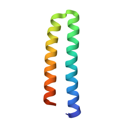
Structure of the ColE1 rop protein at 1.7 A resolution

Banner, D.W.Kokkinidis, M.Tsernoglou, D.

(1987) J Mol Biology 196: 657-675

  • DOI: https://doi.org/10.1016/0022-2836(87)90039-8
  • Primary Citation Related Structures: 
    1ROP

  • PubMed Abstract: 

    Structural details of the Rop protein from plasmid ColE1 are presented, with a description of the X-ray crystal structure determination and refinement at a nominal resolution of 1.7 A. The 63 amino acid protein is a dimer. Each monomer consists almost entirely of two alpha helices, the whole molecule forming a highly regular four-alpha-helix bundle. This may be approximated by a four-stranded rope with a radius of 7.0 A, a left-handed helical twist and a pitch of 172.5 A. The packing constraints for this novel type of coiled-coil structure are given. The protein acts in the control of plasmid replication via regulation of an RNA-RNA interaction in a manner not yet understood in atomic detail.


  • Organizational Affiliation
    • EMBL, Heidelberg, FRG.

Macromolecule Content 

  • Total Structure Weight: 7.24 kDa 
  • Atom Count: 495 
  • Modeled Residue Count: 56 
  • Deposited Residue Count: 63 
  • Unique protein chains: 1

Macromolecules

Find similar proteins by:|  3D Structure
Entity ID: 1
MoleculeChains  Sequence LengthOrganismDetailsImage
ROP PROTEIN63Escherichia coliMutation(s): 0 
UniProt
Find proteins for P03051 (Escherichia coli)
Explore P03051 
Go to UniProtKB:  P03051
Entity Groups
Sequence Clusters30% Identity50% Identity70% Identity90% Identity95% Identity100% Identity
UniProt GroupP03051
Sequence Annotations
Expand
Reference Sequence

Experimental Data & Validation

Experimental Data

  • Method: X-RAY DIFFRACTION
  • Resolution: 1.70 Å
  • R-Value Observed: 0.182 (Depositor) 
Space Group: C 1 2 1
Unit Cell:
Length ( Å )Angle ( ˚ )
a = 60.38α = 90
b = 40.39β = 105.77
c = 27.73γ = 90
Software Package:
Software NamePurpose
PROLSQrefinement

Structure Validation

View Full Validation Report


Entry History 

Deposition Data

Revision History  (Full details and data files)

  • Version 1.0: 1992-07-15
    Type: Initial release
  • Version 1.1: 2008-03-24
    Changes: Version format compliance
  • Version 1.2: 2011-07-13
    Changes: Derived calculations, Version format compliance
  • Version 1.3: 2024-02-14
    Changes: Data collection, Database references, Other