1RMD | pdb_00001rmd

RAG1 DIMERIZATION DOMAIN


Experimental Data Snapshot

  • Method: X-RAY DIFFRACTION
  • Resolution: 2.10 Å
  • R-Value Free: 
    0.257 (Depositor), 0.255 (DCC) 
  • R-Value Work: 
    0.209 (Depositor), 0.214 (DCC) 
  • R-Value Observed: 
    0.209 (Depositor) 

wwPDB Validation 3D Report Full Report

Validation slider image for 1RMD

This is version 1.3 of the entry. See complete history

Literature

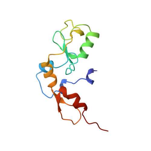
Crystal structure of the RAG1 dimerization domain reveals multiple zinc-binding motifs including a novel zinc binuclear cluster.

Bellon, S.F.Rodgers, K.K.Schatz, D.G.Coleman, J.E.Steitz, T.A.

(1997) Nat Struct Biol 4: 586-591

  • DOI: https://doi.org/10.1038/nsb0797-586
  • Primary Citation Related Structures: 
    1RMD

  • PubMed Abstract: 

    The crystal structure of the dimerization domain of the V(D)J recombination-activating protein, RAG1, was solved using zinc anomalous scattering. The structure reveals an unusual combination of multi-class zinc-binding motifs, including a zinc RING finger and a C2H2 zinc finger, that together from a single structural domain. The domain also contains a unique zinc binuclear cluster in place of a normally mononuclear zinc site in the RING finger. Together, four zinc ions help organize the entire domain, including the two helices that form the dimer interface.


  • Organizational Affiliation
    • Department of Molecular Biophysics and Biochemistry, Yale University, New Haven, Connecticut 06520-8114, USA.

Macromolecule Content 

  • Total Structure Weight: 13.41 kDa 
  • Atom Count: 967 
  • Modeled Residue Count: 116 
  • Deposited Residue Count: 116 
  • Unique protein chains: 1

Macromolecules

Find similar proteins by:|  3D Structure
Entity ID: 1
MoleculeChains  Sequence LengthOrganismDetailsImage
RAG1116Mus musculusMutation(s): 0 
EC: 3.1 (UniProt), 2.3.2.27 (UniProt)
UniProt
Find proteins for P15919 (Mus musculus)
Explore P15919 
Go to UniProtKB:  P15919
Entity Groups
Sequence Clusters30% Identity50% Identity70% Identity90% Identity95% Identity100% Identity
UniProt GroupP15919
Sequence Annotations
Expand
Reference Sequence

Experimental Data & Validation

Experimental Data

  • Method: X-RAY DIFFRACTION
  • Resolution: 2.10 Å
  • R-Value Free:  0.257 (Depositor), 0.255 (DCC) 
  • R-Value Work:  0.209 (Depositor), 0.214 (DCC) 
  • R-Value Observed: 0.209 (Depositor) 
Space Group: P 32 2 1
Unit Cell:
Length ( Å )Angle ( ˚ )
a = 57.58α = 90
b = 57.58β = 90
c = 98.43γ = 120
Software Package:
Software NamePurpose
BAZFAZmodel building
X-PLORrefinement
DENZOdata reduction
SCALEPACKdata scaling
BAZFAZphasing

Structure Validation

View Full Validation Report


Entry History 

Deposition Data

Revision History  (Full details and data files)

  • Version 1.0: 1997-07-23
    Type: Initial release
  • Version 1.1: 2008-03-24
    Changes: Version format compliance
  • Version 1.2: 2011-07-13
    Changes: Version format compliance
  • Version 1.3: 2024-02-14
    Changes: Data collection, Database references, Derived calculations, Other, Structure summary