1RKT | pdb_00001rkt

Crystal structure of yfiR, a putative transcriptional regulator from Bacillus subtilis


Experimental Data Snapshot

  • Method: X-RAY DIFFRACTION
  • Resolution: 1.95 Å
  • R-Value Free: 
    0.254 (Depositor) 
  • R-Value Work: 
    0.208 (Depositor), 0.251 (DCC) 
  • R-Value Observed: 
    0.210 (Depositor) 

wwPDB Validation 3D Report Full Report

Validation slider image for 1RKT

This is version 1.3 of the entry. See complete history

Literature

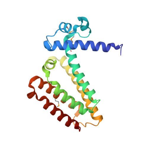
Crystal structure of YfiR, an unusual TetR/CamR-type putative transcriptional regulator from Bacillus subtilis.

Rajan, S.S.Yang, X.Shuvalova, L.Collart, F.Anderson, W.F.

(2006) Proteins 65: 255-257

Macromolecule Content 

  • Total Structure Weight: 47.81 kDa 
  • Atom Count: 3,548 
  • Modeled Residue Count: 404 
  • Deposited Residue Count: 410 
  • Unique protein chains: 1

Macromolecules

Find similar proteins by:|  3D Structure
Entity ID: 1
MoleculeChains  Sequence LengthOrganismDetailsImage
protein yfiR
A, B
205Bacillus subtilisMutation(s): 4 
Gene Names: yfiR
UniProt
Find proteins for O31560 (Bacillus subtilis (strain 168))
Explore O31560 
Go to UniProtKB:  O31560
Entity Groups
Sequence Clusters30% Identity50% Identity70% Identity90% Identity95% Identity100% Identity
UniProt GroupO31560
Sequence Annotations
Expand
Reference Sequence

Small Molecules

Ligands 1 Unique
IDChains Name / Formula / InChI Key2D Diagram3D Interactions
UNX

Query on UNX


Download:Ideal Coordinates CCD File
AA [auth B]
BA [auth B]
C [auth A]
CA [auth B]
D [auth A]
AA [auth B],
BA [auth B],
C [auth A],
CA [auth B],
D [auth A],
DA [auth B],
E [auth A],
EA [auth B],
F [auth A],
FA [auth B],
G [auth A],
GA [auth B],
H [auth A],
HA [auth B],
I [auth A],
IA [auth B],
J [auth A],
JA [auth B],
K [auth A],
KA [auth B],
L [auth A],
LA [auth B],
M [auth A],
MA [auth B],
N [auth A],
NA [auth B],
O [auth A],
OA [auth B],
P [auth A],
Q [auth A],
R [auth A],
S [auth B],
T [auth B],
U [auth B],
V [auth B],
W [auth B],
X [auth B],
Y [auth B],
Z [auth B]
UNKNOWN ATOM OR ION
X
Modified Residues  1 Unique
IDChains TypeFormula2D DiagramParent
MSE
Query on MSE
A, B
L-PEPTIDE LINKINGC5 H11 N O2 SeMET

Experimental Data & Validation

Experimental Data

  • Method: X-RAY DIFFRACTION
  • Resolution: 1.95 Å
  • R-Value Free:  0.254 (Depositor) 
  • R-Value Work:  0.208 (Depositor), 0.251 (DCC) 
  • R-Value Observed: 0.210 (Depositor) 
Space Group: P 1
Unit Cell:
Length ( Å )Angle ( ˚ )
a = 47.983α = 71.96
b = 48.765β = 70.65
c = 57.233γ = 80.2
Software Package:
Software NamePurpose
REFMACrefinement
HKL-2000data reduction
SCALEPACKdata scaling
SOLVEphasing
RESOLVEphasing

Structure Validation

View Full Validation Report


Entry History 

Deposition Data

Revision History  (Full details and data files)

  • Version 1.0: 2004-04-13
    Type: Initial release
  • Version 1.1: 2008-04-29
    Changes: Version format compliance
  • Version 1.2: 2011-07-13
    Changes: Advisory, Version format compliance
  • Version 1.3: 2024-11-13
    Changes: Data collection, Database references, Derived calculations, Refinement description, Structure summary