1RGJ | pdb_00001rgj

NMR STRUCTURE OF THE COMPLEX BETWEEN ALPHA-BUNGAROTOXIN AND MIMOTOPE OF THE NICOTINIC ACETYLCHOLINE RECEPTOR WITH ENHANCED ACTIVITY


Experimental Data Snapshot

  • Method: SOLUTION NMR
  • Conformers Calculated: 100 
  • Conformers Submitted: 
  • Selection Criteria: structures with favorable non-bond energy 

wwPDB Validation 3D Report Full Report

Validation slider image for 1RGJ

This is version 1.4 of the entry. See complete history

Literature

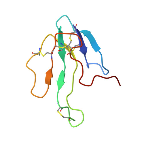
NMR and MD studies on the interaction between ligand peptides and alpha-bungarotoxin.

Bernini, A.Ciutti, A.Spiga, O.Scarselli, M.Klein, S.Vannetti, S.Bracci, L.Lozzi, L.Lelli, B.Falciani, C.Neri, P.Niccolai, N.

(2004) J Mol Biology 339: 1169-1177

  • DOI: https://doi.org/10.1016/j.jmb.2004.04.041
  • Primary Citation Related Structures: 
    1RGJ

  • PubMed Abstract: 

    The interaction between alpha-bungarotoxin and linear synthetic peptides, mimotope of the nicotinic acetylcholine receptor binding site, has been characterised extensively by several methods and a wealth of functional, kinetic and structural data are available. Hence, this system represents a suitable model to explore in detail the dynamics of a peptide-protein interaction. Here, the solution structure of a new complex of the protein toxin with a tridecapeptide ligand exhibiting high affinity has been determined by NMR. As observed for three other previously reported mimotope-alpha-bungarotoxin complexes, also in this case correlations between biological activity and kinetic data are not fully consistent with a static discussion of structural data. Molecular dynamics simulations of the four mimotope-toxin complexes indicate that a relevant contribution to the complex stability is given by the extent of the residual flexibility that the protein maintains upon peptide binding. This feature, limiting the entropy loss caused by protein folding and binding, ought to be generally considered in a rational design of specific protein ligands.


  • Organizational Affiliation
    • Biomolecular Structure Research Center and Department of Molecular Biology, University of Siena, via Fiorentina 1, I-53100 Siena, Italy. andrea.bernini@unisi.it

Macromolecule Content 

  • Total Structure Weight: 9.71 kDa 
  • Atom Count: 671 
  • Modeled Residue Count: 87 
  • Deposited Residue Count: 87 
  • Unique protein chains: 2

Macromolecules

Find similar proteins by:|  3D Structure
Entity ID: 1
MoleculeChains  Sequence LengthOrganismDetailsImage
long neurotoxin 174Bungarus multicinctusMutation(s): 0 
UniProt
Find proteins for P60615 (Bungarus multicinctus)
Explore P60615 
Go to UniProtKB:  P60615
Entity Groups
Sequence Clusters30% Identity50% Identity70% Identity90% Identity95% Identity100% Identity
UniProt GroupP60615
Sequence Annotations
Expand
Reference Sequence
Find similar proteins by:  Sequence   |   3D Structure  
Entity ID: 2
MoleculeChains  Sequence LengthOrganismDetailsImage
MIMOTOPE OF THE NICOTINIC ACETYLCHOLINE RECEPTOR13N/AMutation(s): 0 

Experimental Data & Validation

Experimental Data

  • Method: SOLUTION NMR
  • Conformers Calculated: 100 
  • Conformers Submitted: 
  • Selection Criteria: structures with favorable non-bond energy 

Structure Validation

View Full Validation Report


Entry History 

Deposition Data

Revision History  (Full details and data files)

  • Version 1.0: 2003-11-25
    Type: Initial release
  • Version 1.1: 2008-04-29
    Changes: Version format compliance
  • Version 1.2: 2011-07-13
    Changes: Version format compliance
  • Version 1.3: 2022-03-02
    Changes: Data collection, Database references, Derived calculations
  • Version 1.4: 2024-11-13
    Changes: Data collection, Structure summary