1RGB | pdb_00001rgb

Phospholipase A2 from Vipera ammodytes meridionalis


Experimental Data Snapshot

  • Method: X-RAY DIFFRACTION
  • Resolution: 3.30 Å
  • R-Value Free: 
    0.286 (Depositor) 
  • R-Value Work: 
    0.234 (Depositor) 
  • R-Value Observed: 
    0.234 (Depositor) 

wwPDB Validation 3D Report Full Report

Validation slider image for 1RGB

This is version 1.4 of the entry. See complete history

Literature

Asp49 phospholipase A(2)-elaidoylamide complex: a new mode of inhibition.

Georgieva, D.N.Rypniewski, W.Gabdoulkhakov, A.Genov, N.Betzel, C.

(2004) Biochem Biophys Res Commun 319: 1314-1321

  • DOI: https://doi.org/10.1016/j.bbrc.2004.05.106
  • Primary Citation Related Structures: 
    1RGB

  • PubMed Abstract: 

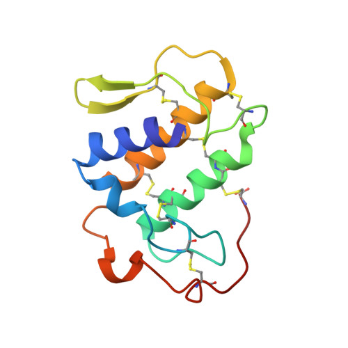
    The inhibition of phospholipase A(2)s (PLA(2)s) is of pharmacological and therapeutic interest because these enzymes are involved in several inflammatory diseases. Elaidoylamide is a powerful inhibitor of a neurotoxic PLA(2) from the Vipera ammodytes meridionalis venom. The X-ray structure of the enzyme-inhibitor complex reveals a new mode of Asp49 PLA(2) inhibition by a fatty acid hydrocarbon chain. The structure contains two identical homodimers in the asymmetric unit. In each dimer one subunit is rotated by 180 degrees with respect to the other and the two molecules are oriented head-to-tail. One molecule of elaidoylamide is bound simultaneously to the substrate binding sites of two associated neurotoxic phospholipase A(2) molecules. The inhibitor binds symmetrically to the hydrophobic channels of the two monomers. The structure can be used to design anti-inflammatory drugs.


  • Organizational Affiliation
    • Institut für Biochemie und Molekularbiologie I, Universitätsklinikum Hamburg-Eppendorf, Hamburg, Germany.

Macromolecule Content 

  • Total Structure Weight: 55.95 kDa 
  • Atom Count: 3,892 
  • Modeled Residue Count: 488 
  • Deposited Residue Count: 488 
  • Unique protein chains: 1

Macromolecules

Find similar proteins by:|  3D Structure
Entity ID: 1
MoleculeChains  Sequence LengthOrganismDetailsImage
Phospholipase A2A,
B,
C [auth K],
D [auth L]
122Vipera ammodytes meridionalisMutation(s): 0 
EC: 3.1.1.4
UniProt
Find proteins for P14420 (Vipera ammodytes meridionalis)
Explore P14420 
Go to UniProtKB:  P14420
Entity Groups
Sequence Clusters30% Identity50% Identity70% Identity90% Identity95% Identity100% Identity
UniProt GroupP14420
Sequence Annotations
Expand
Reference Sequence

Small Molecules

Ligands 1 Unique
IDChains Name / Formula / InChI Key2D Diagram3D Interactions
ELD

Query on ELD


Download:Ideal Coordinates CCD File
E [auth B],
F [auth L]
(9E)-OCTADEC-9-ENAMIDE
C18 H35 N O
FATBGEAMYMYZAF-UHFFFAOYSA-N

Experimental Data & Validation

Experimental Data

  • Method: X-RAY DIFFRACTION
  • Resolution: 3.30 Å
  • R-Value Free:  0.286 (Depositor) 
  • R-Value Work:  0.234 (Depositor) 
  • R-Value Observed: 0.234 (Depositor) 
Space Group: P 21 21 21
Unit Cell:
Length ( Å )Angle ( ˚ )
a = 46.568α = 90
b = 82.67β = 90
c = 119.469γ = 90
Software Package:
Software NamePurpose
CNSrefinement
DENZOdata reduction
SCALEPACKdata scaling
AMoREphasing

Structure Validation

View Full Validation Report


Entry History 

Deposition Data

Revision History  (Full details and data files)

  • Version 1.0: 2005-01-18
    Type: Initial release
  • Version 1.1: 2008-04-29
    Changes: Version format compliance
  • Version 1.2: 2011-07-13
    Changes: Version format compliance
  • Version 1.3: 2018-10-03
    Changes: Advisory, Data collection
  • Version 1.4: 2024-10-30
    Changes: Advisory, Data collection, Database references, Derived calculations, Structure summary