1RF0 | pdb_00001rf0

Crystal Structure of Fragment D of gammaE132A Fibrinogen


Experimental Data Snapshot

  • Method: X-RAY DIFFRACTION
  • Resolution: 2.81 Å
  • R-Value Free: 
    0.286 (Depositor), 0.285 (DCC) 
  • R-Value Work: 
    0.233 (Depositor), 0.233 (DCC) 
  • R-Value Observed: 
    0.233 (Depositor) 

Starting Model: experimental
View more details

wwPDB Validation 3D Report Full Report

Validation slider image for 1RF0

Ligand Structure Quality Assessment 


This is version 1.6 of the entry. See complete history

Literature

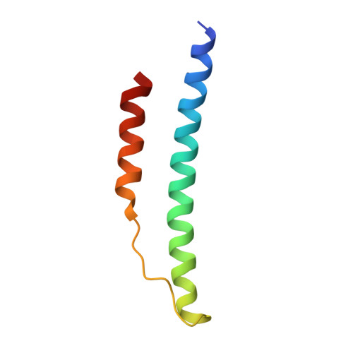
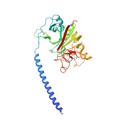
Calcium-Binding Site beta2, Adjacent to the "b" Polymerization Site, Modulates Lateral Aggregation of Protofibrils during Fibrin Polymerization.

Kostelansky, M.S.Lounes, K.C.Ping, L.F.Dickerson, S.K.Gorkun, O.V.Lord, S.T.

(2004) Biochemistry 43: 2475-2483

  • DOI: https://doi.org/10.1021/bi0359978
  • Primary Citation Related Structures: 
    1RF0, 1RF1

  • PubMed Abstract: 

    Structural analysis of recombinant fibrinogen fragment D revealed that the calcium-binding site (beta2-site) composed of residues BbetaAsp261, BbetaAsp398, BbetaGly263, and gammaGlu132 is modulated by the "B:b" interaction. To determine the beta2-site's role in polymerization, we engineered variant fibrinogen gammaE132A in which calcium binding to the beta2-site was disrupted by replacing glutamic acid at gamma132 with alanine. We compared polymerization of gammaE132A to normal fibrinogen as a function of calcium concentration. Polymerization of gammaE132A at concentrations of calcium


  • Organizational Affiliation
    • Department of Chemistry, University of North Carolina, Chapel Hill, North Carolina 27599-7525, USA.

Macromolecule Content 

  • Total Structure Weight: 158.38 kDa 
  • Atom Count: 10,772 
  • Modeled Residue Count: 1,323 
  • Deposited Residue Count: 1,380 
  • Unique protein chains: 3

Macromolecules

Find similar proteins by:|  3D Structure
Entity ID: 1
MoleculeChains  Sequence LengthOrganismDetailsImage
Fibrinogen alpha/alpha-E chain
A, D
66Homo sapiensMutation(s): 0 
Gene Names: FGA
UniProt & NIH Common Fund Data Resources
Find proteins for P02671 (Homo sapiens)
Explore P02671 
Go to UniProtKB:  P02671
PHAROS:  P02671
GTEx:  ENSG00000171560 
Entity Groups
Sequence Clusters30% Identity50% Identity70% Identity90% Identity95% Identity100% Identity
UniProt GroupP02671
Sequence Annotations
Expand
Reference Sequence
Find similar proteins by:|  3D Structure
Entity ID: 2
MoleculeChains  Sequence LengthOrganismDetailsImage
Fibrinogen beta chain
B, E
313Homo sapiensMutation(s): 0 
Gene Names: FGB
UniProt & NIH Common Fund Data Resources
Find proteins for P02675 (Homo sapiens)
Explore P02675 
Go to UniProtKB:  P02675
PHAROS:  P02675
GTEx:  ENSG00000171564 
Entity Groups
Sequence Clusters30% Identity50% Identity70% Identity90% Identity95% Identity100% Identity
UniProt GroupP02675
Glycosylation
Glycosylation Sites: 1Go to GlyGen: P02675-1
Sequence Annotations
Expand
Reference Sequence
Find similar proteins by:|  3D Structure
Entity ID: 3
MoleculeChains  Sequence LengthOrganismDetailsImage
Fibrinogen gamma chain
C, F
311Homo sapiensMutation(s): 1 
Gene Names: FGG
UniProt & NIH Common Fund Data Resources
Find proteins for P02679 (Homo sapiens)
Explore P02679 
Go to UniProtKB:  P02679
PHAROS:  P02679
GTEx:  ENSG00000171557 
Entity Groups
Sequence Clusters30% Identity50% Identity70% Identity90% Identity95% Identity100% Identity
UniProt GroupP02679
Sequence Annotations
Expand
Reference Sequence

Experimental Data & Validation

Experimental Data

  • Method: X-RAY DIFFRACTION
  • Resolution: 2.81 Å
  • R-Value Free:  0.286 (Depositor), 0.285 (DCC) 
  • R-Value Work:  0.233 (Depositor), 0.233 (DCC) 
  • R-Value Observed: 0.233 (Depositor) 
Space Group: P 21 21 21
Unit Cell:
Length ( Å )Angle ( ˚ )
a = 88.408α = 90
b = 94.948β = 90
c = 227.614γ = 90
Software Package:
Software NamePurpose
CNSrefinement
HKL-2000data reduction
SCALEPACKdata scaling
CNSphasing

Structure Validation

View Full Validation Report


Ligand Structure Quality Assessment 


Entry History 

Deposition Data

Revision History  (Full details and data files)

  • Version 1.0: 2004-03-16
    Type: Initial release
  • Version 1.1: 2008-04-29
    Changes: Version format compliance
  • Version 1.2: 2011-07-13
    Changes: Non-polymer description, Version format compliance
  • Version 1.3: 2020-07-29
    Type: Remediation
    Reason: Carbohydrate remediation
    Changes: Data collection, Derived calculations, Structure summary
  • Version 1.4: 2021-10-27
    Changes: Database references, Structure summary
  • Version 1.5: 2023-08-23
    Changes: Data collection, Refinement description
  • Version 1.6: 2024-11-13
    Changes: Structure summary