1RC8 | pdb_00001rc8

T4 Polynucleotide Kinase bound to 5'-GTCAC-3' ssDNA


Experimental Data Snapshot

  • Method: X-RAY DIFFRACTION
  • Resolution: 2.75 Å
  • R-Value Free: 
    0.294 (Depositor), 0.293 (DCC) 
  • R-Value Work: 
    0.230 (Depositor), 0.231 (DCC) 
  • R-Value Observed: 
    0.230 (Depositor) 

Starting Model: experimental
View more details

wwPDB Validation 3D Report Full Report

Validation slider image for 1RC8

Ligand Structure Quality Assessment 


This is version 1.5 of the entry. See complete history

Literature

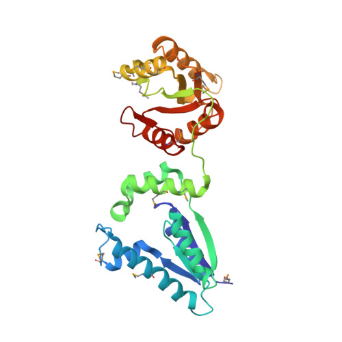
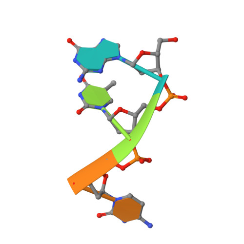
Recognition of DNA substrates by T4 bacteriophage polynucleotide kinase.

Eastberg, J.H.Pelletier, J.Stoddard, B.L.

(2004) Nucleic Acids Res 32: 653-660

  • DOI: https://doi.org/10.1093/nar/gkh212
  • Primary Citation Related Structures: 
    1RC8, 1RPZ, 1RRC

  • PubMed Abstract: 

    T4 phage polynucleotide kinase (PNK) displays 5'-hydroxyl kinase, 3'-phosphatase and 2',3'-cyclic phosphodiesterase activities. The enzyme phosphorylates the 5' hydroxyl termini of a wide variety of nucleic acid substrates, a behavior studied here through the determination of a series of crystal structures with single-stranded (ss)DNA oligonucleotide substrates of various lengths and sequences. In these structures, the 5' ribose hydroxyl is buried in the kinase active site in proper alignment for phosphoryl transfer. Depending on the ssDNA length, the first two or three nucleotide bases are well ordered. Numerous contacts are made both to the phosphoribosyl backbone and to the ordered bases. The position, side chain contacts and internucleotide stacking interactions of the ordered bases are strikingly different for a 5'-GT DNA end than for a 5'-TG end. The base preferences displayed at those positions by PNK are attributable to differences in the enzyme binding interactions and in the DNA conformation for each unique substrate molecule.


  • Organizational Affiliation
    • Fred Hutchinson Cancer Research Center and the Graduate Program in Molecular and Cellular Biology, University of Washington, Seattle, WA 98109, USA.

Macromolecule Content 

  • Total Structure Weight: 37.26 kDa 
  • Atom Count: 2,436 
  • Modeled Residue Count: 292 
  • Deposited Residue Count: 306 
  • Unique protein chains: 1
  • Unique nucleic acid chains: 1

Macromolecules


Find similar proteins by:|  3D Structure
Entity ID: 2
MoleculeChains  Sequence LengthOrganismDetailsImage
Polynucleotide kinaseB [auth A]301Tequatrovirus T4Mutation(s): 12 
Gene Names: PSET
EC: 2.7.1.78 (PDB Primary Data), 3.1.3.34 (UniProt)
UniProt
Find proteins for P06855 (Enterobacteria phage T4)
Explore P06855 
Go to UniProtKB:  P06855
Entity Groups
Sequence Clusters30% Identity50% Identity70% Identity90% Identity95% Identity100% Identity
UniProt GroupP06855
Sequence Annotations
Expand
Reference Sequence
Find similar nucleic acids by:  Sequence
Entity ID: 1
MoleculeChains LengthOrganismImage
5'-D(*GP*TP*CP*AP*C)-3'A [auth B]5N/A
Sequence Annotations
Expand
Reference Sequence

Small Molecules

Modified Residues  1 Unique
IDChains TypeFormula2D DiagramParent
MSE
Query on MSE
B [auth A]L-PEPTIDE LINKINGC5 H11 N O2 SeMET

Experimental Data & Validation

Experimental Data

  • Method: X-RAY DIFFRACTION
  • Resolution: 2.75 Å
  • R-Value Free:  0.294 (Depositor), 0.293 (DCC) 
  • R-Value Work:  0.230 (Depositor), 0.231 (DCC) 
  • R-Value Observed: 0.230 (Depositor) 
Space Group: I 2 2 2
Unit Cell:
Length ( Å )Angle ( ˚ )
a = 82.17α = 90
b = 92.061β = 90
c = 123.385γ = 90
Software Package:
Software NamePurpose
CNSrefinement
HKL-2000data reduction
SCALEPACKdata scaling
EPMRphasing

Structure Validation

View Full Validation Report


Ligand Structure Quality Assessment 


Entry History 

Deposition Data

Revision History  (Full details and data files)

  • Version 1.0: 2004-02-17
    Type: Initial release
  • Version 1.1: 2008-04-29
    Changes: Version format compliance
  • Version 1.2: 2011-07-13
    Changes: Version format compliance
  • Version 1.3: 2023-08-23
    Changes: Data collection, Database references, Derived calculations, Refinement description
  • Version 1.4: 2023-11-15
    Changes: Data collection
  • Version 1.5: 2024-11-13
    Changes: Structure summary