The crystal structure of ribonuclease B at 2.5-A resolution.
Williams, R.L., Greene, S.M., McPherson, A.(1987) J Biological Chem 262: 16020-16031
- PubMed: 3680242 Search on PubMed
- DOI: https://doi.org/10.2210/pdb1rbb/pdb
- Primary Citation Related Structures:
1RBB - PubMed Abstract:
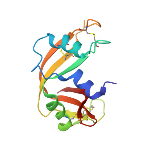
The glycosylated form of bovine pancreatic ribonuclease, RNase B, was crystallized from polyethylene glycol 4000 at low ionic strength in space group C2 with unit cell dimensions of a = 101.81 A, b = 33.36 A, c = 73.60 A, and beta = 90.4 degrees. The crystals, which contained two independent molecules of RNase B as the asymmetric unit, were solved by a combination of multiple isomorphous replacement and molecular replacement approaches. The structures of the two molecules were refined to 2.5-A resolution and a conventional R factor of 0.22 using a constrained-restrained least squares procedure (CORELS). Complexes were also investigated of RNase B plus ruthenium pentaamine and between RNase B and a substrate analogue iodouridine. The polypeptide backbones of the two molecules of RNase B in the asymmetric unit were found to be statistically identical and their differences from RNase A to be statistically insignificant. The carbohydrate chains of both molecules extended into solvent cavities in the crystal lattice and appear to be disordered for the most part. The oligosaccharides appear to exert no influence on the structure of the protein. Iodouridine was observed to bind identically in the pyrimidine site of both RNase B molecules and in a way apparently the same as that previously observed for RNase A. Ruthenium pentaamine bound at histidine 105 of both RNase B molecules in the asymmetric unit, but at a number of secondary sites as well. An array of bound ions was observed by Fo-Fc difference Fourier syntheses. These ions were proximal to lysine and arginine residues at the surface of the proteins while a pair of strong ion binding sites were seen to fall exactly in the active site clefts of both RNase B molecules in the asymmetric unit.
- Department of Biochemistry, University of California, Riverside 92521.
Organizational Affiliation: