1R87 | pdb_00001r87

Crystal structure of the extracellular xylanase from Geobacillus stearothermophilus T-6 (XT6, monoclinic form): The complex of the WT enzyme with xylopentaose at 1.67A resolution


Experimental Data Snapshot

  • Method: X-RAY DIFFRACTION
  • Resolution: 1.67 Å
  • R-Value Free: 
    0.186 (Depositor) 
  • R-Value Work: 
    0.169 (Depositor) 
  • R-Value Observed: 
    0.169 (Depositor) 

Starting Model: experimental
View more details

wwPDB Validation 3D Report Full Report

Currently 1R87 does not have a validation slider image.


This is version 2.1 of the entry. See complete history

Literature

Mapping glycoside hydrolase substrate subsites by isothermal titration calorimetry.

Zolotnitsky, G.Cogan, U.Adir, N.Solomon, V.Shoham, G.Shoham, Y.

(2004) Proc Natl Acad Sci U S A 101: 11275-11280

  • DOI: https://doi.org/10.1073/pnas.0404311101
  • Primary Citation Related Structures: 
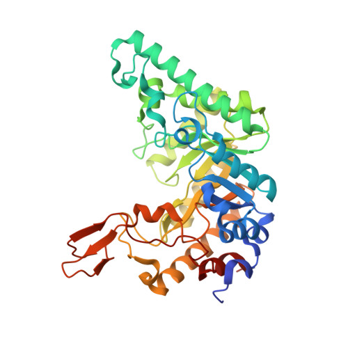
    1R85, 1R87

  • PubMed Abstract: 

    Relating thermodynamic parameters to structural and biochemical data allows a better understanding of substrate binding and its contribution to catalysis. The analysis of the binding of carbohydrates to proteins or enzymes is a special challenge because of the multiple interactions and forces involved. Isothermal titration calorimetry (ITC) provides a direct measure of binding enthalpy (DeltaHa) and allows the determination of the binding constant (free energy), entropy, and stoichiometry. In this study, we used ITC to elucidate the binding thermodynamics of xylosaccharides for two xylanases of family 10 isolated from Geobacillus stearothermophilus T-6. The change in the heat capacity of binding (DeltaCp = DeltaH/DeltaT) for xylosaccharides differing in one sugar unit was determined by using ITC measurements at different temperatures. Because hydrophobic stacking interactions are associated with negative DeltaCp, the data allow us to predict the substrate binding preference in the binding subsites based on the crystal structure of the enzyme. The proposed positional binding preference was consistent with mutants lacking aromatic binding residues at different subsites and was also supported by tryptophan fluorescence analysis.


  • Organizational Affiliation
    • Department of Biotechnology, Technion-Israel Institute of Technology, Haifa 32000, Israel.

Macromolecule Content 

  • Total Structure Weight: 45.19 kDa 
  • Atom Count: 3,451 
  • Modeled Residue Count: 371 
  • Deposited Residue Count: 379 
  • Unique protein chains: 1

Macromolecules

Find similar proteins by:|  3D Structure
Entity ID: 1
MoleculeChains  Sequence LengthOrganismDetailsImage
Endo-1,4-beta-xylanase379Geobacillus stearothermophilusMutation(s): 0 
Gene Names: xynA
EC: 3.2.1.8
UniProt
Find proteins for P40943 (Geobacillus stearothermophilus)
Explore P40943 
Go to UniProtKB:  P40943
Entity Groups
Sequence Clusters30% Identity50% Identity70% Identity90% Identity95% Identity100% Identity
UniProt GroupP40943
Sequence Annotations
Expand
Reference Sequence

Oligosaccharides

Help  
Entity ID: 2
MoleculeChains Length2D Diagram GlycosylationD Interactions
beta-D-xylopyranose-(1-4)-beta-D-xylopyranose
B
2N/A
Glycosylation Resources
GlyTouCan: G87728WL
GlyCosmos: G87728WL
Entity ID: 3
MoleculeChains Length2D Diagram GlycosylationD Interactions
beta-D-xylopyranose-(1-4)-beta-D-xylopyranose-(1-4)-beta-D-xylopyranose
C
3N/A
Glycosylation Resources
GlyTouCan: G87429QT
GlyCosmos: G87429QT

Small Molecules

Ligands 3 Unique
IDChains Name / Formula / InChI Key2D Diagram3D Interactions
SO4

Query on SO4


Download:Ideal Coordinates CCD File
M [auth A]SULFATE ION
O4 S
QAOWNCQODCNURD-UHFFFAOYSA-L
ZN

Query on ZN


Download:Ideal Coordinates CCD File
D [auth A]
E [auth A]
F [auth A]
G [auth A]
H [auth A]
D [auth A],
E [auth A],
F [auth A],
G [auth A],
H [auth A],
I [auth A],
J [auth A]
ZINC ION
Zn
PTFCDOFLOPIGGS-UHFFFAOYSA-N
CL

Query on CL


Download:Ideal Coordinates CCD File
K [auth A],
L [auth A]
CHLORIDE ION
Cl
VEXZGXHMUGYJMC-UHFFFAOYSA-M

Biologically Interesting Molecules (External Reference) 

2 Unique

Experimental Data & Validation

Experimental Data

  • Method: X-RAY DIFFRACTION
  • Resolution: 1.67 Å
  • R-Value Free:  0.186 (Depositor) 
  • R-Value Work:  0.169 (Depositor) 
  • R-Value Observed: 0.169 (Depositor) 
Space Group: C 1 2 1
Unit Cell:
Length ( Å )Angle ( ˚ )
a = 120.422α = 90
b = 61.221β = 119.5
c = 89.583γ = 90
Software Package:
Software NamePurpose
CNSrefinement
DENZOdata reduction
SCALEPACKdata scaling
CNSphasing

Structure Validation

View Full Validation Report

Currently 1R87 does not have a validation slider image.


Entry History 

Deposition Data

Revision History  (Full details and data files)

  • Version 1.0: 2004-07-20
    Type: Initial release
  • Version 1.1: 2008-04-29
    Changes: Version format compliance
  • Version 1.2: 2011-07-13
    Changes: Version format compliance
  • Version 1.3: 2011-11-16
    Changes: Atomic model, Non-polymer description
  • Version 2.0: 2020-07-29
    Type: Remediation
    Reason: Carbohydrate remediation
    Changes: Atomic model, Data collection, Derived calculations, Structure summary
  • Version 2.1: 2023-08-23
    Changes: Data collection, Database references, Derived calculations, Refinement description, Structure summary