1R3N | pdb_00001r3n

Crystal structure of beta-alanine synthase from Saccharomyces kluyveri


Experimental Data Snapshot

  • Method: X-RAY DIFFRACTION
  • Resolution: 2.70 Å
  • R-Value Free: 
    0.266 (Depositor), 0.322 (DCC) 
  • R-Value Work: 
    0.208 (Depositor), 0.292 (DCC) 
  • R-Value Observed: 
    0.211 (Depositor) 

Starting Model: experimental
View more details

wwPDB Validation 3D Report Full Report

Validation slider image for 1R3N

This is version 1.4 of the entry. See complete history

Literature

Yeast beta-Alanine Synthase Shares a Structural Scaffold and Origin with Dizinc-dependent Exopeptidases

Lundgren, S.Gojkovic, Z.Piskur, J.Dobritzsch, D.

(2003) J Biological Chem 278: 51851-51862

  • DOI: https://doi.org/10.1074/jbc.M308674200
  • Primary Citation Related Structures: 
    1R3N, 1R43

  • PubMed Abstract: 

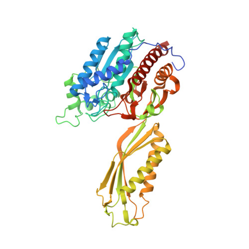
    beta-Alanine synthase (beta AS) is the final enzyme of the reductive pyrimidine catabolic pathway, which is responsible for the breakdown of pyrimidine bases, including several anticancer drugs. In eukaryotes, beta ASs belong to two subfamilies, which exhibit a low degree of sequence similarity. We determined the structure of beta AS from Saccharomyces kluyveri to a resolution of 2.7 A. The subunit of the homodimeric enzyme consists of two domains: a larger catalytic domain with a dizinc metal center, which represents the active site of beta AS, and a smaller domain mediating the majority of the intersubunit contacts. Both domains exhibit a mixed alpha/beta-topology. Surprisingly, the observed high structural homology to a family of dizinc-dependent exopeptidases suggests that these two enzyme groups have a common origin. Alterations in the ligand composition of the metal-binding site can be explained as adjustments to the catalysis of a different reaction, the hydrolysis of an N-carbamyl bond by beta AS compared with the hydrolysis of a peptide bond by exopeptidases. In contrast, there is no resemblance to the three-dimensional structure of the functionally closely related N-carbamyl-d-amino acid amidohydrolases. Based on comparative structural analysis and observed deviations in the backbone conformations of the eight copies of the subunit in the asymmetric unit, we suggest that conformational changes occur during each catalytic cycle.


  • Organizational Affiliation
    • Division of Molecular Structural Biology, Department of Medical Biochemistry and Biophysics, Karolinska Institutet, S-17177 Stockholm, Sweden.

Macromolecule Content 

  • Total Structure Weight: 407.69 kDa 
  • Atom Count: 27,487 
  • Modeled Residue Count: 3,485 
  • Deposited Residue Count: 3,696 
  • Unique protein chains: 1

Macromolecules

Find similar proteins by:|  3D Structure
Entity ID: 1
MoleculeChains  Sequence LengthOrganismDetailsImage
beta-alanine synthase
A, B, C, D, E
A, B, C, D, E, F, G, H
462Lachancea kluyveriMutation(s): 0 
Gene Names: PYD3
EC: 3.5.1.6
UniProt
Find proteins for Q96W94 (Lachancea kluyveri)
Explore Q96W94 
Go to UniProtKB:  Q96W94
Entity Groups
Sequence Clusters30% Identity50% Identity70% Identity90% Identity95% Identity100% Identity
UniProt GroupQ96W94
Sequence Annotations
Expand
Reference Sequence

Small Molecules

Ligands 2 Unique
IDChains Name / Formula / InChI Key2D Diagram3D Interactions
BIB

Query on BIB


Download:Ideal Coordinates CCD File
CA [auth G]
DA [auth G]
K [auth A]
N [auth B]
Q [auth C]
CA [auth G],
DA [auth G],
K [auth A],
N [auth B],
Q [auth C],
T [auth D],
W [auth E],
X [auth E]
BETA-AMINO ISOBUTYRATE
C4 H8 N O2
QCHPKSFMDHPSNR-VKHMYHEASA-M
ZN

Query on ZN


Download:Ideal Coordinates CCD File
AA [auth G]
BA [auth G]
EA [auth H]
FA [auth H]
I [auth A]
AA [auth G],
BA [auth G],
EA [auth H],
FA [auth H],
I [auth A],
J [auth A],
L [auth B],
M [auth B],
O [auth C],
P [auth C],
R [auth D],
S [auth D],
U [auth E],
V [auth E],
Y [auth F],
Z [auth F]
ZINC ION
Zn
PTFCDOFLOPIGGS-UHFFFAOYSA-N

Experimental Data & Validation

Experimental Data

  • Method: X-RAY DIFFRACTION
  • Resolution: 2.70 Å
  • R-Value Free:  0.266 (Depositor), 0.322 (DCC) 
  • R-Value Work:  0.208 (Depositor), 0.292 (DCC) 
  • R-Value Observed: 0.211 (Depositor) 
Space Group: P 1 21 1
Unit Cell:
Length ( Å )Angle ( ˚ )
a = 117.23α = 90
b = 77.12β = 95.05
c = 225.52γ = 90
Software Package:
Software NamePurpose
REFMACrefinement
MOSFLMdata reduction
CCP4data scaling
EPMRphasing

Structure Validation

View Full Validation Report


Entry History 

Deposition Data

Revision History  (Full details and data files)

  • Version 1.0: 2003-11-11
    Type: Initial release
  • Version 1.1: 2008-04-29
    Changes: Version format compliance
  • Version 1.2: 2011-07-13
    Changes: Version format compliance
  • Version 1.3: 2024-02-14
    Changes: Data collection, Database references, Derived calculations, Refinement description
  • Version 1.4: 2024-04-03
    Changes: Refinement description