1R1Z | pdb_00001r1z

The Crystal structure of the Carbohydrate recognition domain of the glycoprotein sorting receptor p58/ERGIC-53 reveals a novel metal binding site and conformational changes associated with calcium ion binding


Experimental Data Snapshot

  • Method: X-RAY DIFFRACTION
  • Resolution: 2.40 Å
  • R-Value Free: 
    0.241 (Depositor), 0.304 (DCC) 
  • R-Value Work: 
    0.223 (Depositor), 0.300 (DCC) 
  • R-Value Observed: 
    0.223 (Depositor) 

Starting Model: experimental
View more details

wwPDB Validation 3D Report Full Report

Validation slider image for 1R1Z

This is version 1.4 of the entry. See complete history

Literature

The Crystal Structure of the Carbohydrate-recognition Domain of the Glycoprotein Sorting Receptor p58/ERGIC-53 Reveals an Unpredicted Metal-binding Site and Conformational Changes Associated with Calcium Ion Binding.

Velloso, L.M.Svensson, K.Pettersson, R.F.Lindqvist, Y.

(2003) J Mol Biology 334: 845-851

  • DOI: https://doi.org/10.1016/j.jmb.2003.10.031
  • Primary Citation Related Structures: 
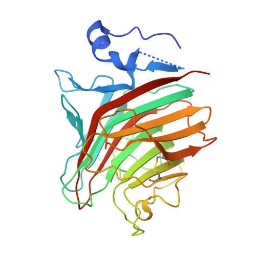
    1R1Z

  • PubMed Abstract: 

    p58/ERGIC-53 is a calcium-dependent animal lectin that acts as a cargo receptor, binding to a set of glycoproteins in the endoplasmic reticulum (ER) and transporting them to the Golgi complex. It is similar in structure to calcium-dependent leguminous lectins. We have determined the structure of the carbohydrate-recognition domain of p58/ERGIC-53 in its calcium-bound form. The structure reveals localized but large conformational changes in relation to the previously determined metal ion-free structure, mapping mostly to the ligand-binding site. It reveals the presence of two calcium ion-binding sites located 6A apart, one of which has no equivalent in the plant lectins. The second metal ion-binding site present in that class of lectins, binding Mn(2+), is absent from p58/ERGIC-53. The absence of a short loop in the ligand-binding site in this protein suggests that it has adapted to optimally bind the high-mannose Man(8)(GlcNAc)(2) glycan common to glycoproteins at the ER exit stage.


  • Organizational Affiliation
    • Molecular Structural Biology, Department of Medical Biochemistry and Biophysics, Karolinska Institutet, S-171 77 Stockholm, Sweden.

Macromolecule Content 

  • Total Structure Weight: 116.43 kDa 
  • Atom Count: 7,793 
  • Modeled Residue Count: 988 
  • Deposited Residue Count: 1,052 
  • Unique protein chains: 1

Macromolecules

Find similar proteins by:|  3D Structure
Entity ID: 1
MoleculeChains  Sequence LengthOrganismDetailsImage
ERGIC-53 protein
A, B, C, D
263Rattus norvegicusMutation(s): 0 
Gene Names: LMAN1 OR ERGIC53
UniProt
Find proteins for Q62902 (Rattus norvegicus)
Explore Q62902 
Go to UniProtKB:  Q62902
Entity Groups
Sequence Clusters30% Identity50% Identity70% Identity90% Identity95% Identity100% Identity
UniProt GroupQ62902
Sequence Annotations
Expand
Reference Sequence

Experimental Data & Validation

Experimental Data

  • Method: X-RAY DIFFRACTION
  • Resolution: 2.40 Å
  • R-Value Free:  0.241 (Depositor), 0.304 (DCC) 
  • R-Value Work:  0.223 (Depositor), 0.300 (DCC) 
  • R-Value Observed: 0.223 (Depositor) 
Space Group: P 1
Unit Cell:
Length ( Å )Angle ( ˚ )
a = 44.351α = 91.05
b = 81.068β = 94.14
c = 82.306γ = 94.99
Software Package:
Software NamePurpose
REFMACrefinement
MOSFLMdata reduction
CCP4data scaling
AMoREphasing

Structure Validation

View Full Validation Report


Entry History 

Deposition Data

Revision History  (Full details and data files)

  • Version 1.0: 2003-12-16
    Type: Initial release
  • Version 1.1: 2008-04-29
    Changes: Version format compliance
  • Version 1.2: 2011-07-13
    Changes: Version format compliance
  • Version 1.3: 2023-08-23
    Changes: Data collection, Database references, Derived calculations, Refinement description
  • Version 1.4: 2024-10-30
    Changes: Structure summary