1QZX | pdb_00001qzx

Crystal structure of the complete core of archaeal SRP and implications for inter-domain communication


Experimental Data Snapshot

  • Method: X-RAY DIFFRACTION
  • Resolution: 4.00 Å
  • R-Value Free: 
    0.383 (Depositor), 0.406 (DCC) 
  • R-Value Work: 
    0.313 (Depositor), 0.409 (DCC) 

Starting Model: experimental
View more details

wwPDB Validation 3D Report Full Report

Validation slider image for 1QZX

This is version 1.3 of the entry. See complete history

Literature

Crystal structure of the complete core of archaeal signal recognition particle and implications for interdomain communication

Rosendal, K.R.Wild, K.Montoya, G.Sinning, I.

(2003) Proc Natl Acad Sci U S A 100: 14701-14706

  • DOI: https://doi.org/10.1073/pnas.2436132100
  • Primary Citation Related Structures: 
    1QZW, 1QZX

  • PubMed Abstract: 

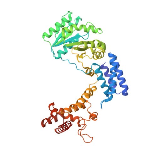
    Targeting of secretory and membrane proteins by the signal recognition particle (SRP) is evolutionarily conserved, and the multidomain protein SRP54 acts as the key player in SRP-mediated protein transport. Binding of a signal peptide to SRP54 at the ribosome is coordinated with GTP binding and subsequent complex formation with the SRP receptor. Because these functions are localized to distinct domains of SRP54, communication between them is essential. We report the crystal structures of SRP54 from the Archaeon Sulfolobus solfataricus with and without its cognate SRP RNA binding site (helix 8) at 4-A resolution. The two structures show the flexibility of the SRP core and the position of SRP54 relative to the RNA. A long linker helix connects the GTPase (G domain) with the signal peptide binding (M) domain, and a hydrophobic contact between the N and M domains relates the signal peptide binding site to the G domain. Hinge regions are identified in the linker between the G and M domains (292-LGMGD) and in the N-terminal part of the M domain, which allow for structural rearrangements within SRP54 upon signal peptide binding at the ribosome.


  • Organizational Affiliation
    • Biochemie-Zentrum Heidelberg, INF 328, D-69120 Heidelberg, Germany.

Macromolecule Content 

  • Total Structure Weight: 98.93 kDa 
  • Atom Count: 6,698 
  • Modeled Residue Count: 850 
  • Deposited Residue Count: 880 
  • Unique protein chains: 1

Macromolecules

Find similar proteins by:|  3D Structure
Entity ID: 1
MoleculeChains  Sequence LengthOrganismDetailsImage
Signal recognition 54 kDa protein
A, B
440Saccharolobus solfataricusMutation(s): 0 
Gene Names: SRP54
EC: 3.6.5.4
UniProt
Find proteins for Q97ZE7 (Saccharolobus solfataricus (strain ATCC 35092 / DSM 1617 / JCM 11322 / P2))
Explore Q97ZE7 
Go to UniProtKB:  Q97ZE7
Entity Groups
Sequence Clusters30% Identity50% Identity70% Identity90% Identity95% Identity100% Identity
UniProt GroupQ97ZE7
Sequence Annotations
Expand
Reference Sequence

Experimental Data & Validation

Experimental Data

  • Method: X-RAY DIFFRACTION
  • Resolution: 4.00 Å
  • R-Value Free:  0.383 (Depositor), 0.406 (DCC) 
  • R-Value Work:  0.313 (Depositor), 0.409 (DCC) 
Space Group: I 4
Unit Cell:
Length ( Å )Angle ( ˚ )
a = 197.906α = 90
b = 197.906β = 90
c = 64.305γ = 90
Software Package:
Software NamePurpose
CNSrefinement
MOSFLMdata reduction
CCP4data scaling
BEASTphasing

Structure Validation

View Full Validation Report


Entry History 

Deposition Data

Revision History  (Full details and data files)

  • Version 1.0: 2003-11-18
    Type: Initial release
  • Version 1.1: 2008-04-29
    Changes: Version format compliance
  • Version 1.2: 2011-07-13
    Changes: Version format compliance
  • Version 1.3: 2023-08-23
    Changes: Data collection, Database references, Derived calculations, Refinement description