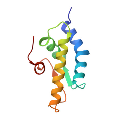
1QZM | pdb_00001qzm

alpha-domain of ATPase


Experimental Data Snapshot

  • Method: X-RAY DIFFRACTION
  • Resolution: 1.90 Å
  • R-Value Free: 
    0.282 (Depositor), 0.267 (DCC) 
  • R-Value Work: 
    0.179 (Depositor), 0.185 (DCC) 
  • R-Value Observed: 
    0.184 (Depositor) 

wwPDB Validation 3D Report Full Report

Validation slider image for 1QZM

This is version 1.4 of the entry. See complete history. 

Literature

Crystal structure of the AAA+ alpha domain of E. coli Lon protease at 1.9A resolution.

Botos, I., Melnikov, E.E., Cherry, S., Khalatova, A.G., Rasulova, F.S., Tropea, J.E., Maurizi, M.R., Rotanova, T.V., Gustchina, A., Wlodawer, A.

(null) J Struct Biol 146: 113-122

  • DOI: https://doi.org/10.1016/j.jsb.2003.09.003
  • Primary Citation Related Structures: 
    1QZM

  • PubMed Abstract: 

    The crystal structure of the small, mostly helical alpha domain of the AAA+ module of the Escherichia coli ATP-dependent protease Lon has been solved by single isomorphous replacement combined with anomalous scattering and refined at 1.9A resolution to a crystallographic R factor of 17.9%. This domain, comprising residues 491-584, was obtained by chymotrypsin digestion of the recombinant full-length protease. The alpha domain of Lon contains four alpha helices and two parallel strands and resembles similar domains found in a variety of ATPases and helicases, including the oligomeric proteases HslVU and ClpAP. The highly conserved "sensor-2" Arg residue is located at the beginning of the third helix. Detailed comparison with the structures of 11 similar domains established the putative location of the nucleotide-binding site in this first fragment of Lon for which a crystal structure has become available.


  • Organizational Affiliation: 
    • Macromolecular Crystallography Laboratory, National Cancer Institute at Frederick, MCL Bldg. 536, Rm. 5, Frederick, MD 21702-1201, USA.

Macromolecule Content 

  • Total Structure Weight: 10.84 kDa 
  • Atom Count: 852 
  • Modeled Residue Count: 94 
  • Deposited Residue Count: 94 
  • Unique protein chains: 1

Macromolecules

Find similar proteins by:|  3D Structure
Entity ID: 1
MoleculeChains  Sequence LengthOrganismDetailsImage
ATP-dependent protease La94Escherichia coliMutation(s): 0 
Gene Names: LON
EC: 3.4.21.53
UniProt
Find proteins for P0A9M0 (Escherichia coli (strain K12))
Explore P0A9M0 
Go to UniProtKB:  P0A9M0
Entity Groups
Sequence Clusters30% Identity50% Identity70% Identity90% Identity95% Identity100% Identity
UniProt GroupP0A9M0
Sequence Annotations
Expand
Reference Sequence

Experimental Data & Validation

Experimental Data

  • Method: X-RAY DIFFRACTION
  • Resolution: 1.90 Å
  • R-Value Free:  0.282 (Depositor), 0.267 (DCC) 
  • R-Value Work:  0.179 (Depositor), 0.185 (DCC) 
  • R-Value Observed: 0.184 (Depositor) 
Space Group: P 43 21 2
Unit Cell:
Length ( Å )Angle ( ˚ )
a = 46.43α = 90
b = 46.43β = 90
c = 85.2γ = 90
Software Package:
Software NamePurpose
SHELXmodel building
SHELXL-97refinement
MAR345data collection
SCALEPACKdata scaling
SHARPphasing
CNSrefinement
SHELXLrefinement

Structure Validation

View Full Validation Report


Entry History 

Deposition Data

Revision History  (Full details and data files)

  • Version 1.0: 2004-05-04
    Type: Initial release
  • Version 1.1: 2007-11-14
    Changes: Version format compliance
  • Version 1.2: 2011-07-13
    Changes: Version format compliance
  • Version 1.3: 2017-10-11
    Changes: Refinement description
  • Version 1.4: 2024-02-14
    Changes: Data collection, Database references