1QSG | pdb_00001qsg

CRYSTAL STRUCTURE OF ENOYL REDUCTASE INHIBITION BY TRICLOSAN


Experimental Data Snapshot

  • Method: X-RAY DIFFRACTION
  • Resolution: 1.75 Å
  • R-Value Free: 
    0.215 (Depositor) 
  • R-Value Work: 
    0.172 (Depositor) 

Starting Model: experimental
View more details

wwPDB Validation 3D Report Full Report

Validation slider image for 1QSG

This is version 1.4 of the entry. See complete history

Literature

Structural basis and mechanism of enoyl reductase inhibition by triclosan.

Stewart, M.J.Parikh, S.Xiao, G.Tonge, P.J.Kisker, C.

(1999) J Mol Biology 290: 859-865

  • DOI: https://doi.org/10.1006/jmbi.1999.2907
  • Primary Citation Related Structures: 
    1QSG

  • PubMed Abstract: 

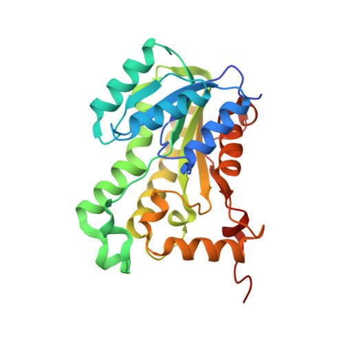
    The enoyl-acyl carrier protein reductase (ENR) is involved in bacterial fatty acid biosynthesis and is the target of the antibacterial diazaborine compounds and the front-line antituberculosis drug isoniazid. Recent studies suggest that ENR is also the target for the broad-spectrum biocide triclosan. The 1.75 A crystal structure of EnvM, the ENR from Escherichia coli, in complex with triclosan and NADH reveals that triclosan binds specifically to EnvM. These data provide a molecular mechanism for the antibacterial activity of triclosan and substantiate the hypothesis that its activity results from inhibition of a specific cellular target rather than non-specific disruption of the bacterial cell membrane. This has important implications for the emergence of drug-resistant bacteria, since triclosan is an additive in many personal care products such as toothpastes, mouthwashes and soaps. Based on this structure, rational design of triclosan derivatives is possible which might be effective against recently identified triclosan-resistant bacterial strains.


  • Organizational Affiliation
    • Department of Pharmacological Sciences, SUNY at Stony Brook, Stony Brook, NY, 11794-8651, USA.

Macromolecule Content 

  • Total Structure Weight: 234.47 kDa 
  • Atom Count: 17,287 
  • Modeled Residue Count: 2,065 
  • Deposited Residue Count: 2,120 
  • Unique protein chains: 1

Macromolecules

Find similar proteins by:|  3D Structure
Entity ID: 1
MoleculeChains  Sequence LengthOrganismDetailsImage
ENOYL-[ACYL-CARRIER-PROTEIN] REDUCTASE
A, B, C, D, E
A, B, C, D, E, F, G, H
265Escherichia coliMutation(s): 0 
EC: 1.3.1.9
UniProt
Find proteins for P0AEK4 (Escherichia coli (strain K12))
Explore P0AEK4 
Go to UniProtKB:  P0AEK4
Entity Groups
Sequence Clusters30% Identity50% Identity70% Identity90% Identity95% Identity100% Identity
UniProt GroupP0AEK4
Sequence Annotations
Expand
Reference Sequence

Small Molecules

Ligands 3 Unique
IDChains Name / Formula / InChI Key2D Diagram3D Interactions
NAD

Query on NAD


Download:Ideal Coordinates CCD File
BA [auth G]
EA [auth H]
J [auth A]
M [auth B]
P [auth C]
BA [auth G],
EA [auth H],
J [auth A],
M [auth B],
P [auth C],
S [auth D],
V [auth E],
Y [auth F]
NICOTINAMIDE-ADENINE-DINUCLEOTIDE
C21 H27 N7 O14 P2
BAWFJGJZGIEFAR-NNYOXOHSSA-N
TCL

Query on TCL


Download:Ideal Coordinates CCD File
CA [auth G]
FA [auth H]
K [auth A]
N [auth B]
Q [auth C]
CA [auth G],
FA [auth H],
K [auth A],
N [auth B],
Q [auth C],
T [auth D],
W [auth E],
Z [auth F]
TRICLOSAN
C12 H7 Cl3 O2
XEFQLINVKFYRCS-UHFFFAOYSA-N
GLC

Query on GLC


Download:Ideal Coordinates CCD File
AA [auth G]
DA [auth H]
I [auth A]
L [auth B]
O [auth C]
AA [auth G],
DA [auth H],
I [auth A],
L [auth B],
O [auth C],
R [auth D],
U [auth E],
X [auth F]
alpha-D-glucopyranose
C6 H12 O6
WQZGKKKJIJFFOK-DVKNGEFBSA-N
Binding Affinity Annotations 
IDSourceBinding Affinity
TCL BindingDB:  1QSG Ki: 7.00e-3 (nM) from 1 assay(s)
IC50: min: 5, max: 900 (nM) from 3 assay(s)

Experimental Data & Validation

Experimental Data

  • Method: X-RAY DIFFRACTION
  • Resolution: 1.75 Å
  • R-Value Free:  0.215 (Depositor) 
  • R-Value Work:  0.172 (Depositor) 
Space Group: P 1
Unit Cell:
Length ( Å )Angle ( ˚ )
a = 73.733α = 89.54
b = 82.077β = 87.43
c = 84.177γ = 77.77
Software Package:
Software NamePurpose
DENZOdata reduction
SCALEPACKdata scaling
AMoREphasing
REFMACrefinement

Structure Validation

View Full Validation Report


Entry History 

Deposition Data

Revision History  (Full details and data files)

  • Version 1.0: 1999-07-21
    Type: Initial release
  • Version 1.1: 2007-10-16
    Changes: Version format compliance
  • Version 1.2: 2011-07-13
    Changes: Derived calculations, Version format compliance
  • Version 1.3: 2020-07-29
    Type: Remediation
    Reason: Carbohydrate remediation
    Changes: Advisory, Data collection, Database references, Derived calculations, Structure summary
  • Version 1.4: 2023-08-16
    Changes: Data collection, Database references, Refinement description, Structure summary