1QRR | pdb_00001qrr

CRYSTAL STRUCTURE OF SQD1 PROTEIN COMPLEX WITH NAD AND UDP-GLUCOSE


Experimental Data Snapshot

  • Method: X-RAY DIFFRACTION
  • Resolution: 1.60 Å
  • R-Value Free: 
    0.191 (Depositor) 
  • R-Value Work: 
    0.170 (Depositor) 
  • R-Value Observed: 
    0.179 (Depositor) 

wwPDB Validation 3D Report Full Report

Validation slider image for 1QRR

This is version 1.4 of the entry. See complete history

Literature

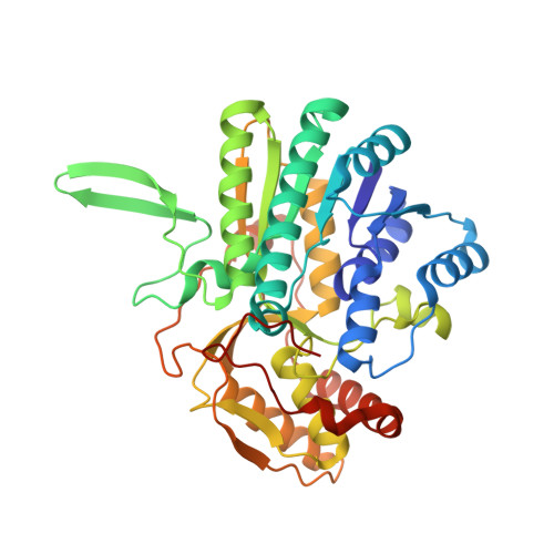
Crystal structure of SQD1, an enzyme involved in the biosynthesis of the plant sulfolipid headgroup donor UDP-sulfoquinovose.

Mulichak, A.M.Theisen, M.J.Essigmann, B.Benning, C.Garavito, R.M.

(1999) Proc Natl Acad Sci U S A 96: 13097-13102

  • DOI: https://doi.org/10.1073/pnas.96.23.13097
  • Primary Citation Related Structures: 
    1QRR

  • PubMed Abstract: 

    The SQD1 enzyme is believed to be involved in the biosynthesis of the sulfoquinovosyl headgroup of plant sulfolipids, catalyzing the transfer of SO(3)(-) to UDP-glucose. We have determined the structure of the complex of SQD1 from Arabidopsis thaliana with NAD(+) and the putative substrate UDP-glucose at 1.6-A resolution. Both bound ligands are completely buried within the binding cleft, along with an internal solvent cavity which is the likely binding site for the, as yet, unidentified sulfur-donor substrate. SQD1 is a member of the short-chain dehydrogenase/reductase (SDR) family of enzymes, and its structure shows a conservation of the SDR catalytic residues. Among several highly conserved catalytic residues, Thr-145 forms unusually short hydrogen bonds with both susceptible hydroxyls of UDP-glucose. A His side chain may also be catalytically important in the sulfonation.


  • Organizational Affiliation
    • Department of Biochemistry, Michigan State University, East Lansing, MI 48824-1319, USA.

Macromolecule Content 

  • Total Structure Weight: 45.79 kDa 
  • Atom Count: 3,481 
  • Modeled Residue Count: 390 
  • Deposited Residue Count: 394 
  • Unique protein chains: 1

Macromolecules

Find similar proteins by:|  3D Structure
Entity ID: 1
MoleculeChains  Sequence LengthOrganismDetailsImage
sulfolipid biosynthesis (SQD1) PROTEIN394Arabidopsis thalianaMutation(s): 0 
EC: 3.13.1.1
UniProt
Find proteins for O48917 (Arabidopsis thaliana)
Explore O48917 
Go to UniProtKB:  O48917
Entity Groups
Sequence Clusters30% Identity50% Identity70% Identity90% Identity95% Identity100% Identity
UniProt GroupO48917
Sequence Annotations
Expand
Reference Sequence

Small Molecules

Ligands 3 Unique
IDChains Name / Formula / InChI Key2D Diagram3D Interactions
NAD

Query on NAD


Download:Ideal Coordinates CCD File
D [auth A]NICOTINAMIDE-ADENINE-DINUCLEOTIDE
C21 H27 N7 O14 P2
BAWFJGJZGIEFAR-NNYOXOHSSA-N
UPG

Query on UPG


Download:Ideal Coordinates CCD File
E [auth A]URIDINE-5'-DIPHOSPHATE-GLUCOSE
C15 H24 N2 O17 P2
HSCJRCZFDFQWRP-JZMIEXBBSA-N
SO4

Query on SO4


Download:Ideal Coordinates CCD File
B [auth A],
C [auth A]
SULFATE ION
O4 S
QAOWNCQODCNURD-UHFFFAOYSA-L

Experimental Data & Validation

Experimental Data

  • Method: X-RAY DIFFRACTION
  • Resolution: 1.60 Å
  • R-Value Free:  0.191 (Depositor) 
  • R-Value Work:  0.170 (Depositor) 
  • R-Value Observed: 0.179 (Depositor) 
Space Group: I 41 2 2
Unit Cell:
Length ( Å )Angle ( ˚ )
a = 159.61α = 90
b = 159.61β = 90
c = 98.89γ = 90
Software Package:
Software NamePurpose
SOLVEphasing
X-PLORrefinement
FRAMBOdata collection
SAINTdata scaling

Structure Validation

View Full Validation Report


Entry History 

Deposition Data

Revision History  (Full details and data files)

  • Version 1.0: 1999-11-10
    Type: Initial release
  • Version 1.1: 2008-04-27
    Changes: Version format compliance
  • Version 1.2: 2011-07-13
    Changes: Version format compliance
  • Version 1.3: 2017-10-04
    Changes: Refinement description
  • Version 1.4: 2024-02-14
    Changes: Data collection, Database references, Derived calculations