1QO0 | pdb_00001qo0

Amide receptor of the amidase operon of Pseudomonas aeruginosa (AmiC) complexed with the negative regulator AmiR.


Experimental Data Snapshot

  • Method: X-RAY DIFFRACTION
  • Resolution: 2.25 Å
  • R-Value Free: 
    0.256 (Depositor), 0.251 (DCC) 
  • R-Value Work: 
    0.186 (Depositor) 

Starting Model: experimental
View more details

wwPDB Validation 3D Report Full Report

Validation slider image for 1QO0

This is version 1.4 of the entry. See complete history

Literature

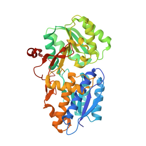
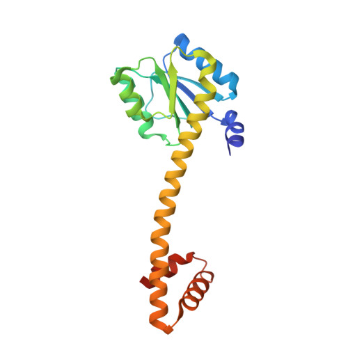
Crystal Structure and Induction Mechanism of Amic-Amir: A Ligand-Regulated Transcription Antitermination Complex

O'Hara, B.P.Norman, R.A.Wan, P.T.C.Roe, S.M.Barrett, T.E.Drew, R.E.Pearl, L.H.

(1999) EMBO J 18: 5175

  • DOI: https://doi.org/10.1093/emboj/18.19.5175
  • Primary Citation Related Structures: 
    1QO0

  • PubMed Abstract: 

    Inducible expression of the aliphatic amidase operon in Pseudomonas aeruginosa is controlled by an antitermination mechanism which allows production of the full-length transcript only in the presence of small-molecule inducers, such as acetamide. Ligand-regulated antitermination is provided by AmiC, the ligand-sensitive negative regulator, and AmiR, the RNA-binding positive regulator. Under non-inducing or repressing growth conditions, AmiC and AmiR form a complex in which the activity of AmiR is silenced. The crystal structure of the AmiC-AmiR complex identifies AmiR as a new and highly unusual member of the response-regulator family of bacterial signal transduction proteins, regulated by sequestration rather than phosphorylation. Comparison with the structure of free AmiC reveals the subtle mechanism of ligand-induced release of AmiR.


  • Organizational Affiliation
    • Centre for Structural Biology, Institute of Cancer Research, Chester Beatty Laboratories, 237 Fulham Road, London SW3 6JB, UK.

Macromolecule Content 

  • Total Structure Weight: 129.9 kDa 
  • Atom Count: 9,661 
  • Modeled Residue Count: 1,130 
  • Deposited Residue Count: 1,162 
  • Unique protein chains: 2

Macromolecules

Find similar proteins by:|  3D Structure
Entity ID: 1
MoleculeChains  Sequence LengthOrganismDetailsImage
AMIC
A, B
385Pseudomonas aeruginosaMutation(s): 0 
UniProt
Find proteins for P27017 (Pseudomonas aeruginosa (strain ATCC 15692 / DSM 22644 / CIP 104116 / JCM 14847 / LMG 12228 / 1C / PRS 101 / PAO1))
Explore P27017 
Go to UniProtKB:  P27017
Entity Groups
Sequence Clusters30% Identity50% Identity70% Identity90% Identity95% Identity100% Identity
UniProt GroupP27017
Sequence Annotations
Expand
Reference Sequence
Find similar proteins by:|  3D Structure
Entity ID: 2
MoleculeChains  Sequence LengthOrganismDetailsImage
AMIRC [auth D],
D [auth E]
196Pseudomonas aeruginosaMutation(s): 0 
UniProt
Find proteins for P10932 (Pseudomonas aeruginosa (strain ATCC 15692 / DSM 22644 / CIP 104116 / JCM 14847 / LMG 12228 / 1C / PRS 101 / PAO1))
Explore P10932 
Go to UniProtKB:  P10932
Entity Groups
Sequence Clusters30% Identity50% Identity70% Identity90% Identity95% Identity100% Identity
UniProt GroupP10932
Sequence Annotations
Expand
Reference Sequence

Experimental Data & Validation

Experimental Data

  • Method: X-RAY DIFFRACTION
  • Resolution: 2.25 Å
  • R-Value Free:  0.256 (Depositor), 0.251 (DCC) 
  • R-Value Work:  0.186 (Depositor) 
Space Group: C 1 2 1
Unit Cell:
Length ( Å )Angle ( ˚ )
a = 308.44α = 90
b = 67.15β = 103.33
c = 76.41γ = 90
Software Package:
Software NamePurpose
REFMACrefinement
MOSFLMdata reduction
SCALAdata scaling
AMoREphasing

Structure Validation

View Full Validation Report


Entry History 

Deposition Data

Revision History  (Full details and data files)

  • Version 1.0: 1999-12-23
    Type: Initial release
  • Version 1.1: 2011-05-08
    Changes: Version format compliance
  • Version 1.2: 2011-07-13
    Changes: Version format compliance
  • Version 1.3: 2019-05-08
    Changes: Data collection, Experimental preparation, Other
  • Version 1.4: 2023-12-13
    Changes: Data collection, Database references, Derived calculations, Other, Refinement description