1QNU | pdb_00001qnu

Shiga-Like Toxin I B Subunit Complexed with the Bridged-Starfish Inhibitor


Experimental Data Snapshot

  • Method: X-RAY DIFFRACTION
  • Resolution: 2.23 Å
  • R-Value Free: 
    0.184 (Depositor), 0.183 (DCC) 
  • R-Value Work: 
    0.171 (Depositor) 
  • R-Value Observed: 
    0.171 (Depositor) 

Starting Model: experimental
View more details

wwPDB Validation 3D Report Full Report

Validation slider image for 1QNU

This is version 2.2 of the entry. See complete history

Literature

Shiga-like toxins are neutralized by tailored multivalent carbohydrate ligands.

Kitov, P.I.Sadowska, J.M.Mulvey, G.Armstrong, G.D.Ling, H.Pannu, N.S.Read, R.J.Bundle, D.R.

(2000) Nature 403: 669-672

  • DOI: https://doi.org/10.1038/35001095
  • Primary Citation Related Structures: 
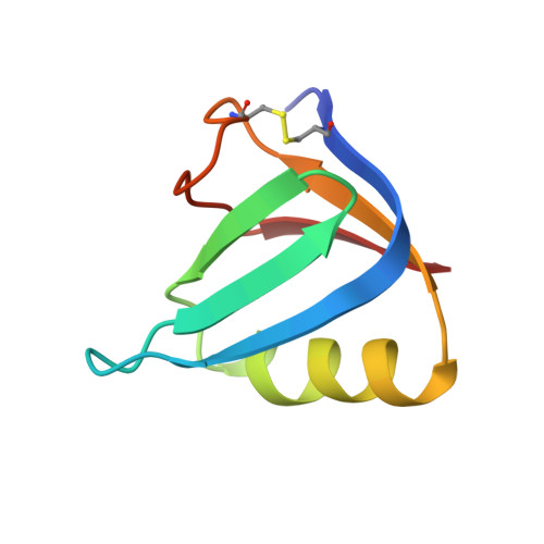
    1QNU

  • PubMed Abstract: 

    The diseases caused by Shiga and cholera toxins account for the loss of millions of lives each year. Both belong to the clinically significant subset of bacterial AB5 toxins consisting of an enzymatically active A subunit that gains entry to susceptible mammalian cells after oligosaccharide recognition by the B5 homopentamer. Therapies might target the obligatory oligosaccharide-toxin recognition event, but the low intrinsic affinity of carbohydrate-protein interactions hampers the development of low-molecular-weight inhibitors. The toxins circumvent low affinity by binding simultaneously to five or more cell-surface carbohydrates. Here we demonstrate the use of the crystal structure of the B5 subunit of Escherichia coli O157:H7 Shiga-like toxin I (SLT-I) in complex with an analogue of its carbohydrate receptor to design an oligovalent, water-soluble carbohydrate ligand (named STARFISH), with subnanomolar inhibitory activity. The in vitro inhibitory activity is 1-10-million-fold higher than that of univalent ligands and is by far the highest molar activity of any inhibitor yet reported for Shiga-like toxins I and II. Crystallography of the STARFISH/Shiga-like toxin I complex explains this activity. Two trisaccharide receptors at the tips of each of five spacer arms simultaneously engage all five B subunits of two toxin molecules.


  • Organizational Affiliation
    • Department of Chemistry, University of Alberta, Edmonton, Canada.

Macromolecule Content 

  • Total Structure Weight: 42.05 kDa 
  • Atom Count: 3,020 
  • Modeled Residue Count: 345 
  • Deposited Residue Count: 345 
  • Unique protein chains: 1

Macromolecules

Find similar proteins by:|  3D Structure
Entity ID: 1
MoleculeChains  Sequence LengthOrganismDetailsImage
Shiga toxin 1 variant B subunit
A, B, C, D, E
69Escherichia coli O157:H7Mutation(s): 0 
Gene Names: stx1
UniProt
Find proteins for V5URS0 (Shigella phage 75/02 Stx)
Explore V5URS0 
Go to UniProtKB:  V5URS0
Entity Groups
Sequence Clusters30% Identity50% Identity70% Identity90% Identity95% Identity100% Identity
UniProt GroupV5URS0
Sequence Annotations
Expand
Reference Sequence

Oligosaccharides

Help  
Entity ID: 2
MoleculeChains Length2D Diagram GlycosylationD Interactions
beta-D-galactopyranose-(1-4)-beta-D-galactopyranose-(1-4)-alpha-D-glucopyranose
F, G, H, I, J
3N/A
Glycosylation Resources
GlyTouCan: G33559CW
GlyCosmos: G33559CW
GlyGen: G33559CW

Small Molecules

Ligands 2 Unique
IDChains Name / Formula / InChI Key2D Diagram3D Interactions
EMB

Query on EMB


Download:Ideal Coordinates CCD File
K [auth A],
M [auth B],
O [auth C],
Q [auth D],
S [auth E]
METHYL-CARBAMIC ACID ETHYL ESTER
C4 H9 N O2
SURZCVYFPAXNGN-UHFFFAOYSA-N
MEC

Query on MEC


Download:Ideal Coordinates CCD File
L [auth A],
N [auth B],
P [auth C],
R [auth D],
T [auth E]
ETHYL-CARBAMIC ACID METHYL ESTER
C4 H9 N O2
AEARPZNULDFPNQ-UHFFFAOYSA-N

Experimental Data & Validation

Experimental Data

  • Method: X-RAY DIFFRACTION
  • Resolution: 2.23 Å
  • R-Value Free:  0.184 (Depositor), 0.183 (DCC) 
  • R-Value Work:  0.171 (Depositor) 
  • R-Value Observed: 0.171 (Depositor) 
Space Group: C 1 2 1
Unit Cell:
Length ( Å )Angle ( ˚ )
a = 104.47α = 90
b = 71.61β = 109.02
c = 56.36γ = 90
Software Package:
Software NamePurpose
CNSrefinement
MOSFLMdata reduction
SCALAdata scaling
CNSphasing

Structure Validation

View Full Validation Report


Entry History 

Deposition Data

Revision History  (Full details and data files)

  • Version 1.0: 2000-04-11
    Type: Initial release
  • Version 1.1: 2011-05-08
    Changes: Version format compliance
  • Version 1.2: 2011-07-13
    Changes: Version format compliance
  • Version 1.3: 2018-05-30
    Changes: Data collection, Structure summary
  • Version 1.4: 2018-06-13
    Changes: Data collection, Database references, Source and taxonomy, Structure summary
  • Version 1.5: 2019-05-08
    Changes: Data collection, Derived calculations, Experimental preparation
  • Version 2.0: 2020-07-29
    Type: Remediation
    Reason: Carbohydrate remediation
    Changes: Advisory, Atomic model, Data collection, Derived calculations, Other, Structure summary
  • Version 2.1: 2023-12-13
    Changes: Data collection, Database references, Derived calculations, Refinement description, Structure summary
  • Version 2.2: 2024-11-06
    Changes: Structure summary