1QNJ | pdb_00001qnj

THE STRUCTURE OF NATIVE PORCINE PANCREATIC ELASTASE AT ATOMIC RESOLUTION (1.1 A)


Experimental Data Snapshot

  • Method: X-RAY DIFFRACTION
  • Resolution: 1.10 Å
  • R-Value Free: 
    0.165 (Depositor), 0.151 (DCC) 
  • R-Value Work: 
    0.120 (DCC) 
  • R-Value Observed: 
    0.127 (Depositor) 

Starting Model: experimental
View more details

wwPDB Validation 3D Report Full Report

Validation slider image for 1QNJ

This is version 1.7 of the entry. See complete history

Literature

Atomic Resolution Structure of Native Porcine Pancreatic Elastase at 1.1 A

Wurtele, M.Hahn, M.Hilpert, K.Hohne, W.

(2000) Acta Crystallogr D Biol Crystallogr 56: 520

  • DOI: https://doi.org/10.1107/s0907444900000299
  • Primary Citation Related Structures: 
    1QNJ

  • PubMed Abstract: 

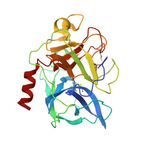
    A data set from the serine protease porcine pancreatic elastase was collected at atomic resolution (1.1 A) with synchrotron radiation. The improved resolution allows the determination of atom positions with high accuracy, as well as the localization of H atoms. Three residues could be modelled in alternative positions. The catalytic triad of elastase consists of His57, Asp102 and Ser195. The His57 N(delta1) H atom was located at a distance of 0.82 A from the N(delta1) atom. The distance between His57 N(delta1) and Asp102 O(delta2) is 2.70 +/- 0.04 A, thus indicating normal hydrogen-bonding geometry. Additional H atoms at His57 N(varepsilon2) and Ser195 O(gamma) could not be identified in the F(o) - F(c) density maps.


  • Organizational Affiliation
    • Institut für Biochemie der Charité, Humboldt-Universität zu Berlin, Monbijoustrasse 2, D-10117 Berlin, Germany.

Macromolecule Content 

  • Total Structure Weight: 26.14 kDa 
  • Atom Count: 2,206 
  • Modeled Residue Count: 240 
  • Deposited Residue Count: 240 
  • Unique protein chains: 1

Macromolecules

Find similar proteins by:|  3D Structure
Entity ID: 1
MoleculeChains  Sequence LengthOrganismDetailsImage
ELASTASE240Sus scrofaMutation(s): 0 
EC: 3.4.21.36
UniProt
Find proteins for P00772 (Sus scrofa)
Explore P00772 
Go to UniProtKB:  P00772
Entity Groups
Sequence Clusters30% Identity50% Identity70% Identity90% Identity95% Identity100% Identity
UniProt GroupP00772
Sequence Annotations
Expand
Reference Sequence

Experimental Data & Validation

Experimental Data

  • Method: X-RAY DIFFRACTION
  • Resolution: 1.10 Å
  • R-Value Free:  0.165 (Depositor), 0.151 (DCC) 
  • R-Value Work:  0.120 (DCC) 
  • R-Value Observed: 0.127 (Depositor) 
Space Group: P 21 21 21
Unit Cell:
Length ( Å )Angle ( ˚ )
a = 49.91α = 90
b = 57.82β = 90
c = 74.27γ = 90
Software Package:
Software NamePurpose
SHELXL-97refinement
DENZOdata reduction
SCALEPACKdata scaling
SHELXL-97phasing

Structure Validation

View Full Validation Report


Entry History 

Deposition Data

Revision History  (Full details and data files)

  • Version 1.0: 2000-03-31
    Type: Initial release
  • Version 1.1: 2011-05-08
    Changes: Version format compliance
  • Version 1.2: 2011-07-13
    Changes: Version format compliance
  • Version 1.3: 2019-05-08
    Changes: Data collection, Experimental preparation, Other
  • Version 1.4: 2019-05-22
    Changes: Data collection, Refinement description
  • Version 1.5: 2019-07-24
    Changes: Data collection
  • Version 1.6: 2023-12-13
    Changes: Data collection, Database references, Derived calculations, Other, Refinement description
  • Version 1.7: 2024-11-13
    Changes: Structure summary