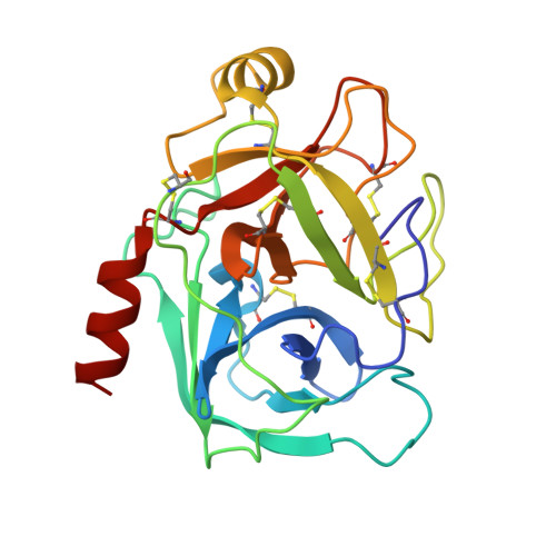
Reconstructing the Binding Site of Factor Xa in Trypsin Reveals Ligand-Induced Structural Plasticity
Reyda, S., Sohn, C., Klebe, G., Rall, K., Ullmann, D., Jakubke, H.D., Stubbs, M.T.(2003) J Mol Biol 325: 963
- PubMed: 12527302
- DOI: https://doi.org/10.1016/s0022-2836(02)01337-2
- Primary Citation of Related Structures:
1J14, 1J15, 1J16, 1J17, 1QL9 - PubMed Abstract:
In order to investigate issues of selectivity and specificity in protein-ligand interactions, we have undertaken the reconstruction of the binding pocket of human factor Xa in the structurally related rat trypsin by site-directed mutagenesis. Three sequential regions (the "99"-, the "175"- and the "190"- loops) were selected as representing the major structural differences between the ligand binding sites of the two enzymes. Wild-type rat trypsin and variants X99rT and X(99/175/190)rT were expressed in yeast, and analysed for their interaction with factor Xa and trypsin inhibitors. For most of the inhibitors studied, progressive loop replacement at the trypsin surface resulted in inhibitory profiles akin to factor Xa. Crystals of the variants were obtained in the presence of benzamidine (3), and could be soaked with the highly specific factor Xa inhibitor (1). Binding of the latter to X99rT results in a series of structural adaptations to the ligand, including the establishment of an "aromatic box" characteristic of factor Xa. In X(99/175/190)rT, introduction of the 175-loop results in a surprising re-orientation of the "intermediate helix", otherwise common to trypsin and factor Xa. The re-orientation is accompanied by an isomerisation of the Cys168-Cys182 disulphide bond, and burial of the critical Phe174 side-chain. In the presence of (1), a major re-organisation of the binding site takes place to yield a geometry identical to that of factor Xa. In all, binding of (1) to trypsin and its variants results in significant structural rearrangements, inducing a binding surface strongly reminiscent of factor Xa, against which the inhibitor was optimised. The structural data reveal a plasticity of the intermediate helix, which has been implicated in the functional cofactor dependency of many trypsin-like serine proteinases. This approach of grafting loops onto scaffolds of known related structures may serve to bridge the gap between structural genomics and drug design.
Organizational Affiliation:
Institut für Pharmazeutische Chemie der Philipps-Universität Marburg, Marbacher Weg 6, D35032, Marburg, Germany.


