1QKI | pdb_00001qki

X-RAY STRUCTURE OF HUMAN GLUCOSE 6-PHOSPHATE DEHYDROGENASE (VARIANT CANTON R459L) COMPLEXED WITH STRUCTURAL NADP+


Experimental Data Snapshot

  • Method: X-RAY DIFFRACTION
  • Resolution: 3.00 Å
  • R-Value Free: 
    0.294 (Depositor) 
  • R-Value Work: 
    0.247 (Depositor), 0.266 (DCC) 
  • R-Value Observed: 
    0.247 (Depositor) 

Starting Model: experimental
View more details

wwPDB Validation 3D Report Full Report

Validation slider image for 1QKI

Ligand Structure Quality Assessment 


This is version 2.1 of the entry. See complete history

Literature

Human Glucose-6-Phosphate Dehydrogenase: The Crystal Structure Reveals a Structural Nadp+ Molecule and Provides Insights Into Enzyme Deficiency

Au, S.W.N.Gover, S.Lam, V.M.S.Adams, M.J.

(2000) Structure 8: 293

  • DOI: https://doi.org/10.1016/s0969-2126(00)00104-0
  • Primary Citation Related Structures: 
    1QKI

  • PubMed Abstract: 

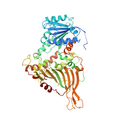
    Glucose-6-phosphate dehydrogenase (G6PD) catalyses the first committed step in the pentose phosphate pathway; the generation of NADPH by this enzyme is essential for protection against oxidative stress. The human enzyme is in a dimer<-->tetramer equilibrium and its stability is dependent on NADP(+) concentration. G6PD deficiency results from many different point mutations in the X-linked gene encoding G6PD and is the most common human enzymopathy. Severe deficiency causes chronic non-spherocytic haemolytic anaemia; the usual symptoms are neonatal jaundice, favism and haemolytic anaemia. We have determined the first crystal structure of a human G6PD (the mutant Canton, Arg459-->Leu) at 3 A resolution. The tetramer is a dimer of dimers. Despite very similar dimer topology, there are two major differences from G6PD of Leuconostoc mesenteroides: a structural NADP(+) molecule, close to the dimer interface but integral to the subunit, is visible in all subunits of the human enzyme; and an intrasubunit disulphide bond tethers the otherwise disordered N-terminal segment. The few dimer-dimer contacts making the tetramer are charge-charge interactions. The importance of NADP(+) for stability is explained by the structural NADP(+) site, which is not conserved in prokaryotes. The structure shows that point mutations causing severe deficiency predominate close to the structural NADP(+) and the dimer interface, primarily affecting the stability of the molecule. They also indicate that a stable dimer is essential to retain activity in vivo. As there is an absolute requirement for some G6PD activity, residues essential for coenzyme or substrate binding are rarely modified.


  • Organizational Affiliation
    • Laboratory of Molecular Biophysics, Department of Biochemistry, University of Oxford, The University of Hong Kong, Department of Biochemistry, Oxford, OX1 3QU, UK, Hong Kong.

Macromolecule Content 

  • Total Structure Weight: 480.36 kDa 
  • Atom Count: 31,663 
  • Modeled Residue Count: 3,912 
  • Deposited Residue Count: 4,112 
  • Unique protein chains: 1

Macromolecules

Find similar proteins by:|  3D Structure
Entity ID: 1
MoleculeChains  Sequence LengthOrganismDetailsImage
GLUCOSE-6-PHOSPHATE 1-DEHYDROGENASE
A, B, C, D, E
A, B, C, D, E, F, G, H
514Homo sapiensMutation(s): 1 
Gene Names: G6PD
EC: 1.1.1.49 (PDB Primary Data), 1 (UniProt)
UniProt & NIH Common Fund Data Resources
Find proteins for P11413 (Homo sapiens)
Explore P11413 
Go to UniProtKB:  P11413
PHAROS:  P11413
GTEx:  ENSG00000160211 
Entity Groups
Sequence Clusters30% Identity50% Identity70% Identity90% Identity95% Identity100% Identity
UniProt GroupP11413
Sequence Annotations
Expand
Reference Sequence

Small Molecules

Ligands 3 Unique
IDChains Name / Formula / InChI Key2D Diagram3D Interactions
NAP

Query on NAP


Download:Ideal Coordinates CCD File
BA [auth H]
I [auth A]
K [auth B]
O [auth C]
R [auth D]
BA [auth H],
I [auth A],
K [auth B],
O [auth C],
R [auth D],
T [auth E],
W [auth F],
Y [auth G]
NADP NICOTINAMIDE-ADENINE-DINUCLEOTIDE PHOSPHATE
C21 H28 N7 O17 P3
XJLXINKUBYWONI-NNYOXOHSSA-N
GOL

Query on GOL


Download:Ideal Coordinates CCD File
AA [auth G],
M [auth B],
N [auth B],
Q [auth C],
V [auth E]
GLYCEROL
C3 H8 O3
PEDCQBHIVMGVHV-UHFFFAOYSA-N
GOA

Query on GOA


Download:Ideal Coordinates CCD File
CA [auth H]
J [auth A]
L [auth B]
P [auth C]
S [auth D]
CA [auth H],
J [auth A],
L [auth B],
P [auth C],
S [auth D],
U [auth E],
X [auth F],
Z [auth G]
GLYCOLIC ACID
C2 H4 O3
AEMRFAOFKBGASW-UHFFFAOYSA-N

Experimental Data & Validation

Experimental Data

  • Method: X-RAY DIFFRACTION
  • Resolution: 3.00 Å
  • R-Value Free:  0.294 (Depositor) 
  • R-Value Work:  0.247 (Depositor), 0.266 (DCC) 
  • R-Value Observed: 0.247 (Depositor) 
Space Group: P 21 21 21
Unit Cell:
Length ( Å )Angle ( ˚ )
a = 128.9α = 90
b = 208.7β = 90
c = 214.3γ = 90
Software Package:
Software NamePurpose
X-PLORrefinement
DENZOdata reduction
SCALAdata scaling
X-PLORphasing

Structure Validation

View Full Validation Report


Ligand Structure Quality Assessment 


Entry History 

Deposition Data

Revision History  (Full details and data files)

  • Version 1.0: 2000-03-16
    Type: Initial release
  • Version 1.1: 2011-05-08
    Changes: Version format compliance
  • Version 1.2: 2011-07-13
    Changes: Version format compliance
  • Version 1.3: 2019-05-08
    Changes: Data collection, Experimental preparation, Other
  • Version 1.4: 2019-05-22
    Changes: Data collection, Refinement description
  • Version 2.0: 2023-11-15
    Changes: Atomic model, Data collection, Database references, Derived calculations, Other
  • Version 2.1: 2023-12-13
    Changes: Refinement description