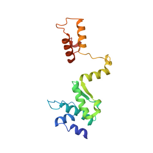
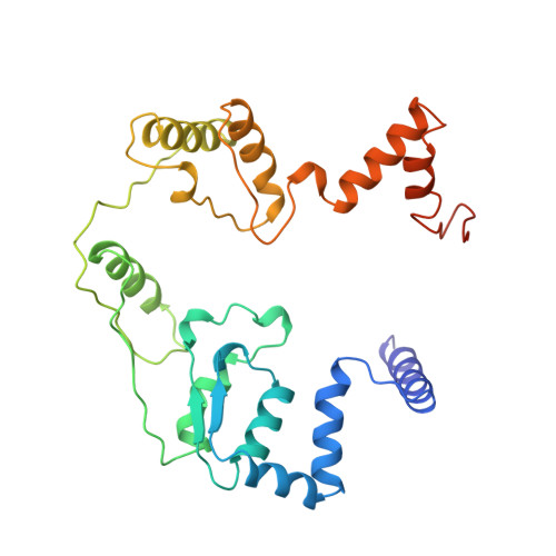
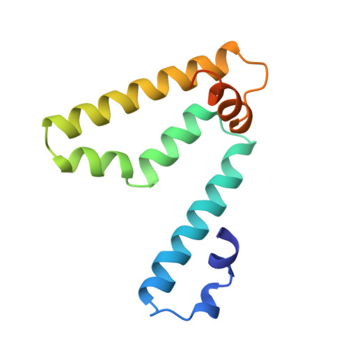
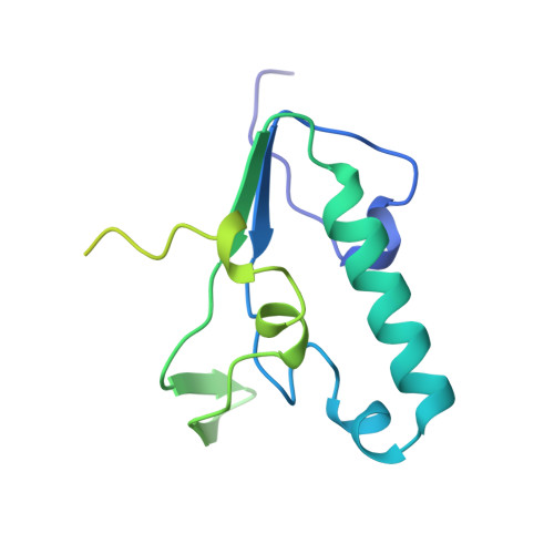
1QHH

STRUCTURE OF DNA HELICASE WITH ADPNP


Experimental Data Snapshot

  • Method: X-RAY DIFFRACTION
  • Resolution: 2.50 Å
  • R-Value Free: 0.285 
  • R-Value Work: 0.228 

wwPDB Validation   3D Report Full Report


Ligand Structure Quality Assessment 


This is version 1.3 of the entry. See complete history


Literature

DNA binding mediates conformational changes and metal ion coordination in the active site of PcrA helicase.

Soultanas, P.Dillingham, M.S.Velankar, S.S.Wigley, D.B.

(1999) J Mol Biol 290: 137-148

  • DOI: https://doi.org/10.1006/jmbi.1999.2873
  • Primary Citation of Related Structures:  
    1QHG, 1QHH

  • PubMed Abstract: 

    Based upon the crystal structures of PcrA helicase, we have made and characterised mutations in a number of conserved helicase signature motifs around the ATPase active site. We have also determined structures of complexes of wild-type PcrA with ADPNP and of a mutant PcrA complexed with ADPNP and Mn2+. The kinetic and structural data define roles for a number of different residues in and around the ATP binding site. More importantly, our results also show that there are two functionally distinct conformations of ATP in the active site. In one conformation, ATP is hydrolysed poorly whereas in the other (activated) conformation, ATP is hydrolysed much more rapidly. We propose a mechanism to explain how the stimulation of ATPase activity afforded by binding of single-stranded DNA stabilises the activated conformation favouring Mg2+binding and a consequent repositioning of the gamma-phosphate group which promotes ATP hydrolysis. A part of the associated conformational change in the protein forces the side-chain of K37 to vacate the Mg2+binding site, allowing the cation to bind and interact with ATP.


  • Organizational Affiliation

    Sir William Dunn School of Pathology, University of Oxford, South Parks Road, Oxford, OX1 3RE, UK.


Macromolecules
Find similar proteins by:  (by identity cutoff)  |  3D Structure
Entity ID: 1
MoleculeChains Sequence LengthOrganismDetailsImage
PROTEIN (PCRA (SUBUNIT))167Geobacillus stearothermophilusMutation(s): 0 
Gene Names: PCRA
EC: 5.6.2.4
UniProt
Find proteins for P56255 (Geobacillus stearothermophilus)
Explore P56255 
Go to UniProtKB:  P56255
Entity Groups  
Sequence Clusters30% Identity50% Identity70% Identity90% Identity95% Identity100% Identity
UniProt GroupP56255
Sequence Annotations
Expand
  • Reference Sequence
Find similar proteins by:  (by identity cutoff)  |  3D Structure
Entity ID: 2
MoleculeChains Sequence LengthOrganismDetailsImage
PROTEIN (PCRA (SUBUNIT))273Geobacillus stearothermophilusMutation(s): 0 
Gene Names: PCRA
EC: 5.6.2.4
UniProt
Find proteins for P56255 (Geobacillus stearothermophilus)
Explore P56255 
Go to UniProtKB:  P56255
Entity Groups  
Sequence Clusters30% Identity50% Identity70% Identity90% Identity95% Identity100% Identity
UniProt GroupP56255
Sequence Annotations
Expand
  • Reference Sequence
Find similar proteins by:  (by identity cutoff)  |  3D Structure
Entity ID: 3
MoleculeChains Sequence LengthOrganismDetailsImage
PROTEIN (PCRA (SUBUNIT))115Geobacillus stearothermophilusMutation(s): 0 
Gene Names: PCRA
EC: 5.6.2.4
UniProt
Find proteins for P56255 (Geobacillus stearothermophilus)
Explore P56255 
Go to UniProtKB:  P56255
Entity Groups  
Sequence Clusters30% Identity50% Identity70% Identity90% Identity95% Identity100% Identity
UniProt GroupP56255
Sequence Annotations
Expand
  • Reference Sequence
Find similar proteins by:  (by identity cutoff)  |  3D Structure
Entity ID: 4
MoleculeChains Sequence LengthOrganismDetailsImage
PROTEIN (PCRA (SUBUNIT))169Geobacillus stearothermophilusMutation(s): 0 
Gene Names: PCRA
EC: 5.6.2.4
UniProt
Find proteins for P56255 (Geobacillus stearothermophilus)
Explore P56255 
Go to UniProtKB:  P56255
Entity Groups  
Sequence Clusters30% Identity50% Identity70% Identity90% Identity95% Identity100% Identity
UniProt GroupP56255
Sequence Annotations
Expand
  • Reference Sequence
Small Molecules
Ligands 1 Unique
IDChains Name / Formula / InChI Key2D Diagram3D Interactions
ATP
Query on ATP

Download Ideal Coordinates CCD File 
E [auth A]ADENOSINE-5'-TRIPHOSPHATE
C10 H16 N5 O13 P3
ZKHQWZAMYRWXGA-KQYNXXCUSA-N
Experimental Data & Validation

Experimental Data

  • Method: X-RAY DIFFRACTION
  • Resolution: 2.50 Å
  • R-Value Free: 0.285 
  • R-Value Work: 0.228 
  • Space Group: P 65
Unit Cell:
Length ( Å )Angle ( ˚ )
a = 138.936α = 90
b = 138.936β = 90
c = 111.642γ = 120
Software Package:
Software NamePurpose
X-PLORrefinement
DENZOdata reduction

Structure Validation

View Full Validation Report


Ligand Structure Quality Assessment 


Entry History 

Deposition Data

Revision History  (Full details and data files)

  • Version 1.0: 1999-07-13
    Type: Initial release
  • Version 1.1: 2008-04-26
    Changes: Version format compliance
  • Version 1.2: 2011-07-13
    Changes: Version format compliance
  • Version 1.3: 2023-12-27
    Changes: Data collection, Database references, Derived calculations