1QH3

HUMAN GLYOXALASE II WITH CACODYLATE AND ACETATE IONS PRESENT IN THE ACTIVE SITE


Experimental Data Snapshot

  • Method: X-RAY DIFFRACTION
  • Resolution: 1.90 Å
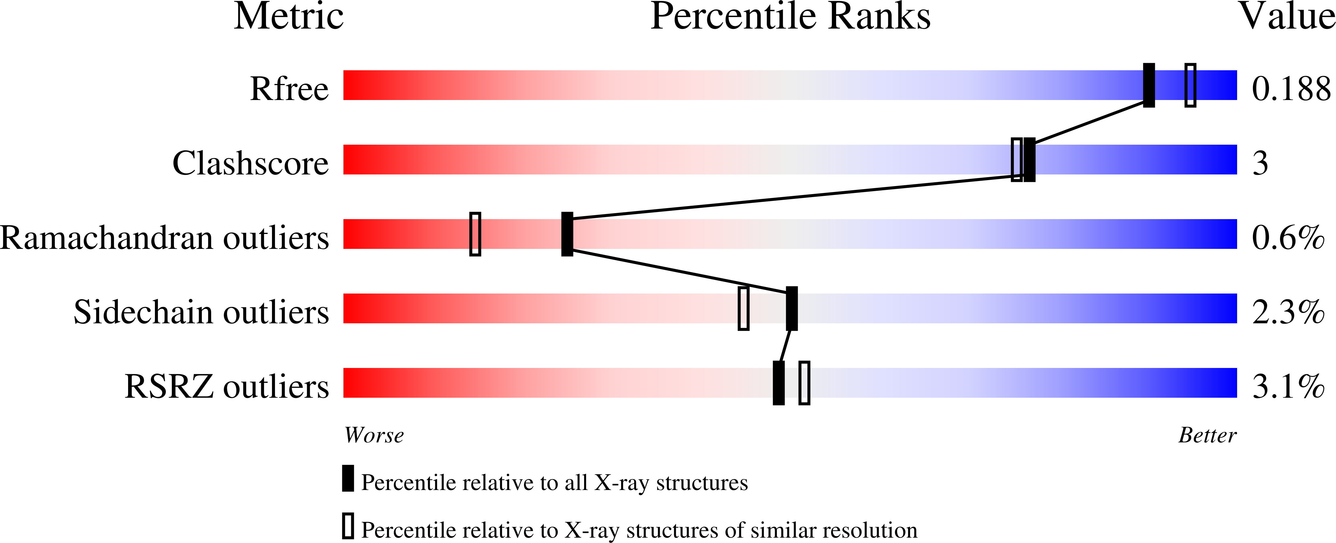
  • R-Value Free: 0.239 
  • R-Value Work: 0.184 
  • R-Value Observed: 0.185 

wwPDB Validation   3D Report Full Report


This is version 1.3 of the entry. See complete history


Literature

Crystal structure of human glyoxalase II and its complex with a glutathione thiolester substrate analogue.

Cameron, A.D.Ridderstrom, M.Olin, B.Mannervik, B.

(1999) Structure 7: 1067-1078

  • DOI: https://doi.org/10.1016/s0969-2126(99)80174-9
  • Primary Citation of Related Structures:  
    1QH3, 1QH5

  • PubMed Abstract: 

    Glyoxalase II, the second of two enzymes in the glyoxalase system, is a thiolesterase that catalyses the hydrolysis of S-D-lactoylglutathione to form glutathione and D-lactic acid. The structure of human glyoxalase II was solved initially by single isomorphous replacement with anomalous scattering and refined at a resolution of 1.9 A. The enzyme consists of two domains. The first domain folds into a four-layered beta sandwich, similar to that seen in the metallo-beta-lactamases. The second domain is predominantly alpha-helical. The active site contains a binuclear zinc-binding site and a substrate-binding site extending over the domain interface. The model contains acetate and cacodylate in the active site. A second complex was derived from crystals soaked in a solution containing the slow substrate, S-(N-hydroxy-N-bromophenylcarbamoyl)glutathione. This complex was refined at a resolution of 1.45 A. It contains the added ligand in one molecule of the asymmetric unit and glutathione in the other. The arrangement of ligands around the zinc ions includes a water molecule, presumably in the form of a hydroxide ion, coordinated to both metal ions. This hydroxide ion is situated 2.9 A from the carbonyl carbon of the substrate in such a position that it could act as the nucleophile during catalysis. The reaction mechanism may also have implications for the action of metallo-beta-lactamases.


  • Organizational Affiliation

    Department of Molecular Biology Uppsala University Biomedical Center Box 590, S-751 24, Uppsala, Sweden Structural Biology Laboratory Department of Chemistry University of York Heslington, York, UK YO10 5DD,. cameron@yorvic.york.ac.uk.


Macromolecules
Find similar proteins by:  (by identity cutoff)  |  3D Structure
Entity ID: 1
MoleculeChains Sequence LengthOrganismDetailsImage
PROTEIN (HYDROXYACYLGLUTATHIONE HYDROLASE)
A, B
260Homo sapiensMutation(s): 0 
EC: 3.1.2.6
UniProt & NIH Common Fund Data Resources
Find proteins for Q16775 (Homo sapiens)
Explore Q16775 
Go to UniProtKB:  Q16775
PHAROS:  Q16775
GTEx:  ENSG00000063854 
Entity Groups  
Sequence Clusters30% Identity50% Identity70% Identity90% Identity95% Identity100% Identity
UniProt GroupQ16775
Sequence Annotations
Expand
  • Reference Sequence
Small Molecules
Ligands 5 Unique
IDChains Name / Formula / InChI Key2D Diagram3D Interactions
CAC
Query on CAC

Download Ideal Coordinates CCD File 
E [auth A],
G [auth A],
L [auth B],
N [auth B]
CACODYLATE ION
C2 H6 As O2
OGGXGZAMXPVRFZ-UHFFFAOYSA-M
ZN
Query on ZN

Download Ideal Coordinates CCD File 
C [auth A],
D [auth A],
J [auth B],
K [auth B]
ZINC ION
Zn
PTFCDOFLOPIGGS-UHFFFAOYSA-N
ACT
Query on ACT

Download Ideal Coordinates CCD File 
F [auth A],
M [auth B]
ACETATE ION
C2 H3 O2
QTBSBXVTEAMEQO-UHFFFAOYSA-M
MN
Query on MN

Download Ideal Coordinates CCD File 
H [auth A],
O [auth B]
MANGANESE (II) ION
Mn
WAEMQWOKJMHJLA-UHFFFAOYSA-N
CL
Query on CL

Download Ideal Coordinates CCD File 
I [auth A]CHLORIDE ION
Cl
VEXZGXHMUGYJMC-UHFFFAOYSA-M
Experimental Data & Validation

Experimental Data

  • Method: X-RAY DIFFRACTION
  • Resolution: 1.90 Å
  • R-Value Free: 0.239 
  • R-Value Work: 0.184 
  • R-Value Observed: 0.185 
  • Space Group: P 21 21 21
Unit Cell:
Length ( Å )Angle ( ˚ )
a = 39.08α = 90
b = 72.37β = 90
c = 162.06γ = 90
Software Package:
Software NamePurpose
SHARPphasing
REFMACrefinement
DENZOdata reduction
SCALEPACKdata scaling

Structure Validation

View Full Validation Report


Entry History 

Deposition Data

Revision History  (Full details and data files)

  • Version 1.0: 1999-09-24
    Type: Initial release
  • Version 1.1: 2007-10-16
    Changes: Version format compliance
  • Version 1.2: 2011-07-13
    Changes: Non-polymer description, Version format compliance
  • Version 1.3: 2023-12-27
    Changes: Data collection, Database references, Derived calculations