1QDR | pdb_00001qdr

2.1 A RESOLUTION STRUCTURE OF ESCHERICHIA COLI LYTIC TRANSGLYCOSYLASE SLT35


Experimental Data Snapshot

  • Method: X-RAY DIFFRACTION
  • Resolution: 2.10 Å
  • R-Value Free: 
    0.210 (Depositor) 
  • R-Value Work: 
    0.158 (Depositor), 0.172 (DCC) 
  • R-Value Observed: 
    0.158 (Depositor) 

wwPDB Validation 3D Report Full Report

Validation slider image for 1QDR

Ligand Structure Quality Assessment 


This is version 1.4 of the entry. See complete history

Literature

Binding of calcium in the EF-hand of Escherichia coli lytic transglycosylase Slt35 is important for stability.

van Asselt, E.J.Dijkstra, B.W.

(1999) FEBS Lett 458: 429-435

  • DOI: https://doi.org/10.1016/s0014-5793(99)01198-9
  • Primary Citation Related Structures: 
    1QDR, 1QDT

  • PubMed Abstract: 

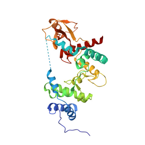
    The Escherichia coli lytic transglycosylase Slt35 contains a single metal ion-binding site that resembles EF-hand calcium-binding sites. The Slt35 EF-hand is only the second observation of such a domain in a prokaryotic protein. Two crystal structures at 2.1 A resolution show that both Ca2+ ions and Na+ ions can bind to the EF-hand domain, but in subtly different configurations. Heat-induced unfolding studies demonstrate that Ca2+ ions are preferentially bound, and that only Ca2+ ions significantly increase the melting temperature of Slt35. This shows that the EF-hand calcium-binding domain is important for the stability of Slt35.


  • Organizational Affiliation
    • BIOSON Research Institute and Laboratory of Biophysical Chemistry, University of Groningen, The Netherlands.

Macromolecule Content 

  • Total Structure Weight: 36.33 kDa 
  • Atom Count: 2,863 
  • Modeled Residue Count: 312 
  • Deposited Residue Count: 322 
  • Unique protein chains: 1

Macromolecules

Find similar proteins by:|  3D Structure
Entity ID: 1
MoleculeChains  Sequence LengthOrganismDetailsImage
LYTIC MUREIN TRANSGLYCOSYLASE B322Escherichia coliMutation(s): 2 
EC: 3.2.1
UniProt
Find proteins for P41052 (Escherichia coli (strain K12))
Explore P41052 
Go to UniProtKB:  P41052
Entity Groups
Sequence Clusters30% Identity50% Identity70% Identity90% Identity95% Identity100% Identity
UniProt GroupP41052
Sequence Annotations
Expand
Reference Sequence

Experimental Data & Validation

Experimental Data

  • Method: X-RAY DIFFRACTION
  • Resolution: 2.10 Å
  • R-Value Free:  0.210 (Depositor) 
  • R-Value Work:  0.158 (Depositor), 0.172 (DCC) 
  • R-Value Observed: 0.158 (Depositor) 
Space Group: P 21 21 21
Unit Cell:
Length ( Å )Angle ( ˚ )
a = 58.533α = 90
b = 67.799β = 90
c = 98.72γ = 90
Software Package:
Software NamePurpose
X-PLORmodel building
X-PLORrefinement
DENZOdata reduction
SCALEPACKdata scaling
X-PLORphasing

Structure Validation

View Full Validation Report


Ligand Structure Quality Assessment 


Entry History 

Deposition Data

Revision History  (Full details and data files)

  • Version 1.0: 2000-01-24
    Type: Initial release
  • Version 1.1: 2008-04-27
    Changes: Version format compliance
  • Version 1.2: 2011-07-13
    Changes: Version format compliance
  • Version 1.3: 2021-11-03
    Changes: Database references, Derived calculations
  • Version 1.4: 2024-02-14
    Changes: Data collection