1QDO | pdb_00001qdo

MAN(APLHA1-3)MAN(ALPHA1-O)METHYL CONCANAVALIN A COMPLEX


Experimental Data Snapshot

  • Method: X-RAY DIFFRACTION
  • Resolution: 2.80 Å
  • R-Value Free: 
    0.257 (Depositor), 0.248 (DCC) 
  • R-Value Work: 
    0.164 (Depositor), 0.166 (DCC) 

Starting Model: experimental
View more details

wwPDB Validation 3D Report Full Report

Validation slider image for 1QDO

This is version 2.1 of the entry. See complete history

Literature

The crystal structures of Man(alpha1-3)Man(alpha1-O)Me and Man(alpha1-6)Man(alpha1-O)Me in complex with concanavalin A.

Bouckaert, J.Hamelryck, T.W.Wyns, L.Loris, R.

(1999) J Biological Chem 274: 29188-29195

  • DOI: https://doi.org/10.1074/jbc.274.41.29188
  • Primary Citation Related Structures: 
    1QDC, 1QDO

  • PubMed Abstract: 

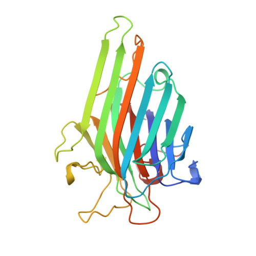
    The crystal structures of concanavalin A in complex with Man(alpha1-6)Man(alpha1-O)Me and Man(alpha1-3)Man(alpha1-O)Me were determined at resolutions of 2.0 and 2.8 A, respectively. In both structures, the O-1-linked mannose binds in the conserved monosaccharide-binding site. The O-3-linked mannose of Man(alpha1-3)Man(alpha1-O)Me binds in the hydrophobic subsite formed by Tyr-12, Tyr-100, and Leu-99. The shielding of a hydrophobic surface is consistent with the associated large heat capacity change. The O-6-linked mannose of Man(alpha1-6)Man(alpha1-O)Me binds in the same subsite formed by Tyr-12 and Asp-16 as the reducing mannose of the highly specific trimannose Man(alpha1-3)[Man(alpha1-6)]Man(alpha1-O)Me. However, it is much less tightly bound. Its O-2 hydroxyl makes no hydrogen bond with the conserved water 1. Water 1 is present in all the sugar-containing concanavalin A structures and increases the complementarity between the protein-binding surface and the sugar, but is not necessarily a hydrogen-bonding partner. A water analysis of the carbohydrate-binding site revealed a conserved water molecule replacing O-4 on the alpha1-3-linked arm of the trimannose. No such water is found for the reducing or O-6-linked mannose. Our data indicate that the central mannose of Man(alpha1-3)[Man(alpha1-6)]Man(alpha1-O)Me primarily functions as a hinge between the two outer subsites.


  • Organizational Affiliation
    • Laboratorium voor Ultrastructuur, Vlaams Interuniversitair Instituut voor Biotechnologie, Vrije Universiteit Brussel, Paardenstraat 65, B-1640 Sint-Genesius-Rode, Belgium. bouckaej@vub.ac.be

Macromolecule Content 

  • Total Structure Weight: 104.29 kDa 
  • Atom Count: 7,373 
  • Modeled Residue Count: 948 
  • Deposited Residue Count: 948 
  • Unique protein chains: 1

Macromolecules

Find similar proteins by:|  3D Structure
Entity ID: 1
MoleculeChains  Sequence LengthOrganismDetailsImage
PROTEIN (CONCANAVALIN A)
A, B, C, D
237Canavalia ensiformisMutation(s): 0 
UniProt
Find proteins for P55915 (Canavalia brasiliensis)
Explore P55915 
Go to UniProtKB:  P55915
Entity Groups
Sequence Clusters30% Identity50% Identity70% Identity90% Identity95% Identity100% Identity
UniProt GroupP55915
Sequence Annotations
Expand
Reference Sequence

Oligosaccharides

Help  
Entity ID: 2
MoleculeChains Length2D Diagram GlycosylationD Interactions
alpha-D-mannopyranose-(1-3)-methyl alpha-D-mannopyranoside
E, F, G, H
2N/A
Glycosylation Resources
GlyTouCan: G59594NN
GlyCosmos: G59594NN

Small Molecules

Ligands 2 Unique
IDChains Name / Formula / InChI Key2D Diagram3D Interactions
MN

Query on MN


Download:Ideal Coordinates CCD File
I [auth A],
K [auth B],
M [auth C],
O [auth D]
MANGANESE (II) ION
Mn
WAEMQWOKJMHJLA-UHFFFAOYSA-N
CA

Query on CA


Download:Ideal Coordinates CCD File
J [auth A],
L [auth B],
N [auth C],
P [auth D]
CALCIUM ION
Ca
BHPQYMZQTOCNFJ-UHFFFAOYSA-N

Experimental Data & Validation

Experimental Data

  • Method: X-RAY DIFFRACTION
  • Resolution: 2.80 Å
  • R-Value Free:  0.257 (Depositor), 0.248 (DCC) 
  • R-Value Work:  0.164 (Depositor), 0.166 (DCC) 
Space Group: P 1 21 1
Unit Cell:
Length ( Å )Angle ( ˚ )
a = 60.41α = 90
b = 64.53β = 93.21
c = 126.96γ = 90
Software Package:
Software NamePurpose
AMoREphasing
X-PLORrefinement
DENZOdata reduction
SCALEPACKdata scaling

Structure Validation

View Full Validation Report


Entry History 

Deposition Data

Revision History  (Full details and data files)

  • Version 1.0: 1999-10-14
    Type: Initial release
  • Version 1.1: 2008-04-27
    Changes: Version format compliance
  • Version 1.2: 2011-07-13
    Changes: Version format compliance
  • Version 2.0: 2020-07-29
    Type: Remediation
    Reason: Carbohydrate remediation
    Changes: Atomic model, Data collection, Derived calculations, Structure summary
  • Version 2.1: 2023-08-16
    Changes: Data collection, Database references, Refinement description, Structure summary