1QAZ | pdb_00001qaz

CRYSTAL STRUCTURE OF ALGINATE LYASE A1-III FROM SPHINGOMONAS SPECIES A1 AT 1.78A RESOLUTION


Experimental Data Snapshot

  • Method: X-RAY DIFFRACTION
  • Resolution: 1.78 Å
  • R-Value Free: 
    0.233 (Depositor) 
  • R-Value Work: 
    0.180 (Depositor), 0.355 (DCC) 

wwPDB Validation 3D Report Full Report

Validation slider image for 1QAZ

This is version 1.4 of the entry. See complete history

Literature

Crystal structure of alginate lyase A1-III from Sphingomonas species A1 at 1.78 A resolution.

Yoon, H.J.Mikami, B.Hashimoto, W.Murata, K.

(1999) J Mol Biology 290: 505-514

  • DOI: https://doi.org/10.1006/jmbi.1999.2883
  • Primary Citation Related Structures: 
    1QAZ

  • PubMed Abstract: 

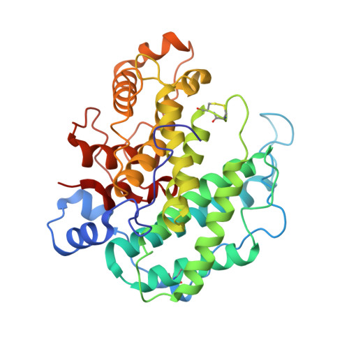
    The three-dimensional structure of alginate lyase A1-III (ALYIII) from a Sphingomonas species A1 was determined by X-ray crystallography. The enzyme was crystallized by the hanging-drop vapour-diffusion method in the presence of 49% ammonium sulfate at 20 degrees C. The crystals are monoclinic and belong to the space group C2 with unit cell dimensions of a=49.18 A, b=93.08 A, c=82.10 A and beta=104.12 degrees. There was one molecule of alginate lyase in the asymmetric unit of the crystal. The diffraction data up to 1. 71 A were collected with Rsymof 5.0%. The crystal structure of ALYIII was solved by the multiple isomorphous replacement method and refined at 1.78 A resolution using X-PLOR with a final R -factor of 18.0% for 10.0 to 1.78 A resolution data. The refined model of ALYIII contained 351 amino acid residues, 299 water molecules and two sulfate ions. The three-dimensional structure of ALYIII was abundant in helices and had a deep tunnel-like cleft in a novel (alpha6/alpha5)-barrel structure, which was similar to the (alpha6/alpha6)-barrel found in glucoamylase and cellulase. This structure presented the possibility that alginate molecules might penetrate into the cleft to interact with the catalytic site of ALYIII.


  • Organizational Affiliation
    • Kyoto University, Uji Kyoto, 611-0011, Japan.

Macromolecule Content 

  • Total Structure Weight: 39.84 kDa 
  • Atom Count: 3,101 
  • Modeled Residue Count: 351 
  • Deposited Residue Count: 351 
  • Unique protein chains: 1

Macromolecules

Find similar proteins by:|  3D Structure
Entity ID: 1
MoleculeChains  Sequence LengthOrganismDetailsImage
PROTEIN (ALGINATE LYASE A1-III)351Sphingomonas sp.Mutation(s): 0 
EC: 3.5.1.45
UniProt
Find proteins for A0ACD6B938 (Sphingomonas sp)
Explore A0ACD6B938 
Go to UniProtKB:  A0ACD6B938
Entity Groups
Sequence Clusters30% Identity50% Identity70% Identity90% Identity95% Identity100% Identity
UniProt GroupA0ACD6B938
Sequence Annotations
Expand
Reference Sequence

Experimental Data & Validation

Experimental Data

  • Method: X-RAY DIFFRACTION
  • Resolution: 1.78 Å
  • R-Value Free:  0.233 (Depositor) 
  • R-Value Work:  0.180 (Depositor), 0.355 (DCC) 
Space Group: C 1 2 1
Unit Cell:
Length ( Å )Angle ( ˚ )
a = 48.9α = 90
b = 92.4β = 104
c = 81.6γ = 90
Software Package:
Software NamePurpose
FRAMBOdata collection
SADIEdata reduction
SAINTdata reduction
PHASESphasing
X-PLORrefinement
SADIEdata scaling
SAINTdata scaling

Structure Validation

View Full Validation Report


Entry History 

Deposition Data

  • Released Date: 1999-07-19 
  • Deposition Author(s): Yoon, H.-J.

Revision History  (Full details and data files)

  • Version 1.0: 1999-07-19
    Type: Initial release
  • Version 1.1: 2008-04-27
    Changes: Version format compliance
  • Version 1.2: 2011-07-13
    Changes: Version format compliance
  • Version 1.3: 2017-10-04
    Changes: Refinement description
  • Version 1.4: 2024-11-06
    Changes: Data collection, Database references, Derived calculations, Structure summary