1Q7E | pdb_00001q7e

Crystal Structure of YfdW protein from E. coli


Experimental Data Snapshot

  • Method: X-RAY DIFFRACTION
  • Resolution: 1.60 Å
  • R-Value Free: 
    0.186 (Depositor), 0.189 (DCC) 
  • R-Value Work: 
    0.156 (Depositor), 0.158 (DCC) 

wwPDB Validation 3D Report Full Report

Validation slider image for 1Q7E

This is version 1.4 of the entry. See complete history

Literature

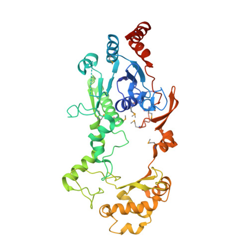
Structure of Escherichia coli YfdW, a type III CoA transferase.

Gogos, A.Gorman, J.Shapiro, L.

(2004) Acta Crystallogr D Biol Crystallogr 60: 507-511

  • DOI: https://doi.org/10.1107/S0907444904000034
  • Primary Citation Related Structures: 
    1PQY, 1Q6Y, 1Q7E

  • PubMed Abstract: 

    Crystal structures are reported for free and coenzyme A (CoA) bound forms of the YfdW protein from Escherichia coli, a representative type III CoA transferase. The structures reveal a two-domain protomer with interdomain connections forming a ring-like structure with a large central hole. Two protomers associate to form a highly intertwined dimer in which the hole of each ring is filled by the partner molecule. Each protomer binds a single CoA molecule and these CoA-binding sites are distant from one another in the dimer.


  • Organizational Affiliation
    • Department of Biochemistry and Molecular Biophysics, Columbia University College of Physicians and Surgeons, 630 West 168th Street, New York, NY 10032, USA.

Macromolecule Content 

  • Total Structure Weight: 48.78 kDa 
  • Atom Count: 3,687 
  • Modeled Residue Count: 410 
  • Deposited Residue Count: 428 
  • Unique protein chains: 1

Macromolecules

Find similar proteins by:|  3D Structure
Entity ID: 1
MoleculeChains  Sequence LengthOrganismDetailsImage
Hypothetical protein yfdW428Escherichia coliMutation(s): 9 
Gene Names: YFDW
EC: 2.8.3.16
UniProt
Find proteins for P69902 (Escherichia coli (strain K12))
Explore P69902 
Go to UniProtKB:  P69902
Entity Groups
Sequence Clusters30% Identity50% Identity70% Identity90% Identity95% Identity100% Identity
UniProt GroupP69902
Sequence Annotations
Expand
Reference Sequence

Small Molecules

Ligands 2 Unique
IDChains Name / Formula / InChI Key2D Diagram3D Interactions
MET

Query on MET


Download:Ideal Coordinates CCD File
B [auth A],
C [auth A]
METHIONINE
C5 H11 N O2 S
FFEARJCKVFRZRR-BYPYZUCNSA-N
MPD

Query on MPD


Download:Ideal Coordinates CCD File
D [auth A]
E [auth A]
F [auth A]
G [auth A]
H [auth A]
D [auth A],
E [auth A],
F [auth A],
G [auth A],
H [auth A],
I [auth A],
J [auth A]
(4S)-2-METHYL-2,4-PENTANEDIOL
C6 H14 O2
SVTBMSDMJJWYQN-YFKPBYRVSA-N
Modified Residues  1 Unique
IDChains TypeFormula2D DiagramParent
MSE
Query on MSE
A
L-PEPTIDE LINKINGC5 H11 N O2 SeMET

Experimental Data & Validation

Experimental Data

  • Method: X-RAY DIFFRACTION
  • Resolution: 1.60 Å
  • R-Value Free:  0.186 (Depositor), 0.189 (DCC) 
  • R-Value Work:  0.156 (Depositor), 0.158 (DCC) 
Space Group: C 1 2 1
Unit Cell:
Length ( Å )Angle ( ˚ )
a = 92.366α = 90
b = 69.605β = 108.63
c = 73.219γ = 90
Software Package:
Software NamePurpose
DENZOdata reduction
SCALEPACKdata scaling
SHARPphasing
REFMACrefinement

Structure Validation

View Full Validation Report


Entry History 

Deposition Data

Revision History  (Full details and data files)

  • Version 1.0: 2003-09-30
    Type: Initial release
  • Version 1.1: 2008-04-29
    Changes: Version format compliance
  • Version 1.2: 2011-07-13
    Changes: Derived calculations, Version format compliance
  • Version 1.3: 2021-02-03
    Changes: Database references, Derived calculations, Structure summary
  • Version 1.4: 2024-11-20
    Changes: Data collection, Database references, Structure summary