1Q1J | pdb_00001q1j

Crystal Structure Analysis of anti-HIV-1 Fab 447-52D in complex with V3 peptide


Experimental Data Snapshot

  • Method: X-RAY DIFFRACTION
  • Resolution: 2.50 Å
  • R-Value Free: 
    0.285 (Depositor), 0.284 (DCC) 
  • R-Value Work: 
    0.250 (Depositor), 0.249 (DCC) 
  • R-Value Observed: 
    0.250 (Depositor) 

Starting Model: experimental
View more details

wwPDB Validation 3D Report Full Report

Validation slider image for 1Q1J

This is version 1.4 of the entry. See complete history

Literature

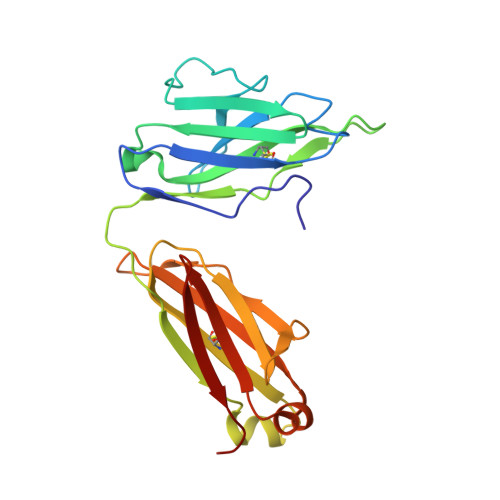
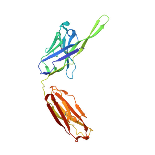
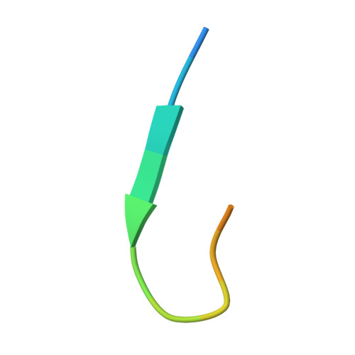
Structural rationale for the broad neutralization of HIV-1 by human monoclonal antibody 447-52D.

Stanfield, R.L.Gorny, M.K.Williams, C.Zolla-Pazner, S.Wilson, I.A.

(2004) Structure 12: 193-204

  • DOI: https://doi.org/10.1016/j.str.2004.01.003
  • Primary Citation Related Structures: 
    1Q1J

  • PubMed Abstract: 

    447-52D is a human monoclonal antibody isolated from a heterohybridoma derived from an HIV-1-infected individual. This antibody recognizes the hypervariable gp120 V3 loop, and neutralizes both X4 and R5 primary isolates, making it one of the most effective anti-V3 antibodies characterized to date. The crystal structure of the 447-52D Fab in complex with a 16-mer V3 peptide at 2.5 A resolution reveals that the peptide beta hairpin forms a three-stranded mixed beta sheet with complementarity determining region (CDR) H3, with most of the V3 side chains exposed to solvent. Sequence specificity is conferred through interaction of the type-II turn (residues GPGR) at the apex of the V3 hairpin with the base of CDR H3. This novel mode of peptide-antibody recognition enables the antibody to bind to many different V3 sequences where only the GPxR core epitope is absolutely required.


  • Organizational Affiliation
    • Department of Molecular Biology, The Scripps Research Institute, 10550 North Torrey Pines Road, La Jolla, CA 92037, USA.

Macromolecule Content 

  • Total Structure Weight: 98.55 kDa 
  • Atom Count: 6,875 
  • Modeled Residue Count: 912 
  • Deposited Residue Count: 924 
  • Unique protein chains: 3

Macromolecules

Find similar proteins by:|  3D Structure
Entity ID: 1
MoleculeChains  Sequence LengthOrganismDetailsImage
Fab 447-52D, light chainA [auth L],
D [auth M]
215Homo sapiensMutation(s): 0 
Find similar proteins by:|  3D Structure
Entity ID: 2
MoleculeChains  Sequence LengthOrganismDetailsImage
Fab 447-52D, heavy chainB [auth H],
E [auth I]
231Homo sapiensMutation(s): 0 
Find similar proteins by:  Sequence   |   3D Structure  
Entity ID: 3
MoleculeChains  Sequence LengthOrganismDetailsImage
gp120 V3 peptideC [auth P],
F [auth Q]
16N/AMutation(s): 0 
UniProt
Find proteins for P05877 (Human immunodeficiency virus type 1 group M subtype B (isolate MN))
Explore P05877 
Go to UniProtKB:  P05877
Entity Groups
Sequence Clusters30% Identity50% Identity70% Identity90% Identity95% Identity100% Identity
UniProt GroupP05877
Sequence Annotations
Expand
Reference Sequence

Experimental Data & Validation

Experimental Data

  • Method: X-RAY DIFFRACTION
  • Resolution: 2.50 Å
  • R-Value Free:  0.285 (Depositor), 0.284 (DCC) 
  • R-Value Work:  0.250 (Depositor), 0.249 (DCC) 
  • R-Value Observed: 0.250 (Depositor) 
Space Group: P 1 21 1
Unit Cell:
Length ( Å )Angle ( ˚ )
a = 76.673α = 90
b = 74.903β = 100.57
c = 100.047γ = 90
Software Package:
Software NamePurpose
HKL-2000data collection
SCALEPACKdata scaling
AMoREphasing
CNSrefinement
HKL-2000data reduction

Structure Validation

View Full Validation Report


Entry History 

Deposition Data

Revision History  (Full details and data files)

  • Version 1.0: 2004-02-17
    Type: Initial release
  • Version 1.1: 2008-04-29
    Changes: Version format compliance
  • Version 1.2: 2011-07-13
    Changes: Version format compliance
  • Version 1.3: 2023-08-16
    Changes: Data collection, Database references, Refinement description
  • Version 1.4: 2024-11-06
    Changes: Structure summary