1Q0H | pdb_00001q0h

Crystal structure of selenomethionine-labelled DXR in complex with fosmidomycin


Experimental Data Snapshot

  • Method: X-RAY DIFFRACTION
  • Resolution: 2.20 Å
  • R-Value Free: 
    0.212 (Depositor), 0.222 (DCC) 
  • R-Value Work: 
    0.187 (Depositor), 0.196 (DCC) 
  • R-Value Observed: 
    0.189 (Depositor) 

Starting Model: experimental
View more details

wwPDB Validation 3D Report Full Report

Validation slider image for 1Q0H

Ligand Structure Quality Assessment 


This is version 1.6 of the entry. See complete history

Literature

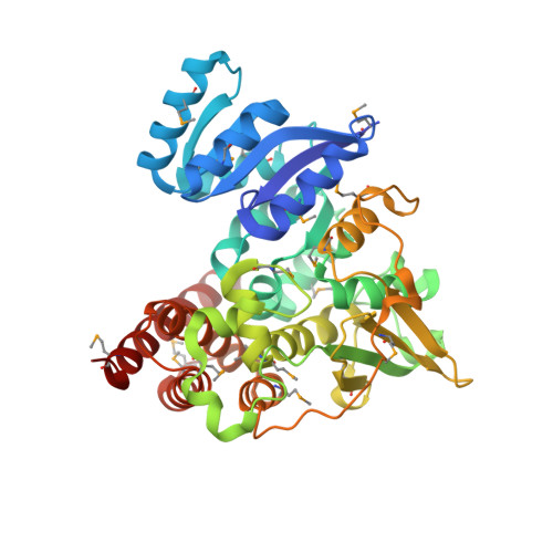
The crystal structure of E.coli 1-deoxy-D-xylulose-5-phosphate reductoisomerase in a ternary complex with the antimalarial compound fosmidomycin and NADPH reveals a tight-binding closed enzyme conformation.

Mac Sweeney, A.Lange, R.Fernandes, R.P.Schulz, H.Dale, G.E.Douangamath, A.Proteau, P.J.Oefner, C.

(2005) J Mol Biology 345: 115-127

  • DOI: https://doi.org/10.1016/j.jmb.2004.10.030
  • Primary Citation Related Structures: 
    1Q0H, 1Q0L, 1Q0Q

  • PubMed Abstract: 

    The key enzyme in the non-mevalonate pathway of isoprenoid biosynthesis, 1-deoxy-D-xylulose 5-phosphate reductoisomerase (DXR) has been shown to be the target enzyme of fosmidomycin, an antimalarial, antibacterial and herbicidal compound. Here we report the crystal structure of selenomethionine-labelled Escherichia coli DXR in a ternary complex with NADPH and fosmidomycin at 2.2 A resolution. The structure reveals a considerable conformational rearrangement upon fosmidomycin binding and provides insights into the slow, tight binding inhibition mode of the inhibitor. Although the inhibitor displays an unusual non-metal mediated mode of inhibition, which is an artefact most likely due to the low metal affinity of DXR at the pH used for crystallization, the structural data add valuable information for the rational design of novel DXR inhibitors. Using this structure together with the published structural data and the 1.9 A crystal structure of DXR in a ternary complex with NADPH and the substrate 1-deoxy-D-xylulose 5-phosphate, a model for the physiologically relevant tight-binding mode of inhibition is proposed. The structure of the substrate complex must be interpreted with caution due to the presence of a second diastereomer in the active site.


  • Organizational Affiliation
    • Morphochem AG, WRO-1055/338, Schwarzwaldallee 215, CH-4058, Basel, Switzerland.

Macromolecule Content 

  • Total Structure Weight: 46.42 kDa 
  • Atom Count: 3,382 
  • Modeled Residue Count: 398 
  • Deposited Residue Count: 406 
  • Unique protein chains: 1

Macromolecules

Find similar proteins by:|  3D Structure
Entity ID: 1
MoleculeChains  Sequence LengthOrganismDetailsImage
1-deoxy-D-xylulose 5-phosphate reductoisomerase406Escherichia coliMutation(s): 19 
Gene Names: DXR
EC: 1.1.1.267
UniProt
Find proteins for P45568 (Escherichia coli (strain K12))
Explore P45568 
Go to UniProtKB:  P45568
Entity Groups
Sequence Clusters30% Identity50% Identity70% Identity90% Identity95% Identity100% Identity
UniProt GroupP45568
Sequence Annotations
Expand
Reference Sequence

Small Molecules

Ligands 3 Unique
IDChains Name / Formula / InChI Key2D Diagram3D Interactions
NDP

Query on NDP


Download:Ideal Coordinates CCD File
C [auth A]NADPH DIHYDRO-NICOTINAMIDE-ADENINE-DINUCLEOTIDE PHOSPHATE
C21 H30 N7 O17 P3
ACFIXJIJDZMPPO-NNYOXOHSSA-N
CIT

Query on CIT


Download:Ideal Coordinates CCD File
D [auth A]CITRIC ACID
C6 H8 O7
KRKNYBCHXYNGOX-UHFFFAOYSA-N
FOM

Query on FOM


Download:Ideal Coordinates CCD File
B [auth A]3-[FORMYL(HYDROXY)AMINO]PROPYLPHOSPHONIC ACID
C4 H10 N O5 P
GJXWDTUCERCKIX-UHFFFAOYSA-N
Modified Residues  1 Unique
IDChains TypeFormula2D DiagramParent
MSE
Query on MSE
A
L-PEPTIDE LINKINGC5 H11 N O2 SeMET
Binding Affinity Annotations 
IDSourceBinding Affinity
FOM BindingDB:  1Q0H IC50: min: 20, max: 438 (nM) from 6 assay(s)

Experimental Data & Validation

Experimental Data

  • Method: X-RAY DIFFRACTION
  • Resolution: 2.20 Å
  • R-Value Free:  0.212 (Depositor), 0.222 (DCC) 
  • R-Value Work:  0.187 (Depositor), 0.196 (DCC) 
  • R-Value Observed: 0.189 (Depositor) 
Space Group: P 31 2 1
Unit Cell:
Length ( Å )Angle ( ˚ )
a = 109.168α = 90
b = 109.168β = 90
c = 90.63γ = 120
Software Package:
Software NamePurpose
DENZOdata reduction
SCALEPACKdata scaling
AMoREphasing
REFMACrefinement

Structure Validation

View Full Validation Report


Ligand Structure Quality Assessment 


Entry History 

Deposition Data

Revision History  (Full details and data files)

  • Version 1.0: 2004-07-20
    Type: Initial release
  • Version 1.1: 2008-04-29
    Changes: Version format compliance
  • Version 1.2: 2011-07-13
    Changes: Derived calculations, Version format compliance
  • Version 1.3: 2018-10-03
    Changes: Advisory, Data collection
  • Version 1.4: 2023-08-16
    Changes: Advisory, Data collection, Database references, Derived calculations, Refinement description
  • Version 1.5: 2023-11-15
    Changes: Data collection
  • Version 1.6: 2024-10-16
    Changes: Structure summary