1PZ3 | pdb_00001pz3

Crystal structure of a family 51 (GH51) alpha-L-arabinofuranosidase from Geobacillus stearothermophilus T6


Experimental Data Snapshot

  • Method: X-RAY DIFFRACTION
  • Resolution: 1.75 Å
  • R-Value Free: 
    0.204 (Depositor), 0.371 (DCC) 
  • R-Value Work: 
    0.171 (Depositor), 0.365 (DCC) 

wwPDB Validation 3D Report Full Report

Validation slider image for 1PZ3

This is version 1.4 of the entry. See complete history

Literature

Crystal structure and snapshots along the reaction pathway of a family 51 alpha-L-arabinofuranosidase

Hoevel, K.Shallom, D.Niefind, K.Belakhov, V.Shoham, G.Baasov, T.Shoham, Y.Schomburg, D.

(2003) EMBO J 22: 4922-4932

  • DOI: https://doi.org/10.1093/emboj/cdg494
  • Primary Citation Related Structures: 
    1PZ2, 1PZ3, 1QW8, 1QW9

  • PubMed Abstract: 

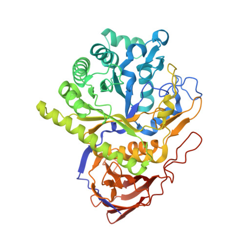
    High-resolution crystal structures of alpha-L-arabinofuranosidase from Geobacillus stearothermophilus T-6, a family 51 glycosidase, are described. The enzyme is a hexamer, and each monomer is organized into two domains: a (beta/alpha)8-barrel and a 12-stranded beta sandwich with jelly-roll topology. The structures of the Michaelis complexes with natural and synthetic substrates, and of the transient covalent arabinofuranosyl-enzyme intermediate represent two stable states in the double displacement mechanism, and allow thorough examination of the catalytic mechanism. The arabinofuranose sugar is tightly bound and distorted by an extensive network of hydrogen bonds. The two catalytic residues are 4.7 A apart, and together with other conserved residues contribute to the stabilization of the oxocarbenium ion-like transition state via charge delocalization and specific protein-substrate interactions. The enzyme is an anti-protonator, and a 1.7 A electrophilic migration of the anomeric carbon takes place during the hydrolysis.


  • Organizational Affiliation
    • Institute for Biochemistry, University of Cologne, Cologne 50674, Germany.

Macromolecule Content 

  • Total Structure Weight: 114.75 kDa 
  • Atom Count: 9,136 
  • Modeled Residue Count: 994 
  • Deposited Residue Count: 1,004 
  • Unique protein chains: 1

Macromolecules

Find similar proteins by:|  3D Structure
Entity ID: 1
MoleculeChains  Sequence LengthOrganismDetailsImage
Alpha-L-arabinofuranosidase
A, B
502Geobacillus stearothermophilusMutation(s): 0 
Gene Names: ABFA
EC: 3.2.1.55
UniProt
Find proteins for Q9XBQ3 (Geobacillus stearothermophilus)
Explore Q9XBQ3 
Go to UniProtKB:  Q9XBQ3
Entity Groups
Sequence Clusters30% Identity50% Identity70% Identity90% Identity95% Identity100% Identity
UniProt GroupQ9XBQ3
Sequence Annotations
Expand
Reference Sequence

Experimental Data & Validation

Experimental Data

  • Method: X-RAY DIFFRACTION
  • Resolution: 1.75 Å
  • R-Value Free:  0.204 (Depositor), 0.371 (DCC) 
  • R-Value Work:  0.171 (Depositor), 0.365 (DCC) 
Space Group: H 3
Unit Cell:
Length ( Å )Angle ( ˚ )
a = 179.43α = 90
b = 179.43β = 90
c = 100.231γ = 120
Software Package:
Software NamePurpose
MAR345data collection
SCALEPACKdata scaling
REFMACrefinement

Structure Validation

View Full Validation Report


Entry History 

Deposition Data

Revision History  (Full details and data files)

  • Version 1.0: 2003-10-07
    Type: Initial release
  • Version 1.1: 2008-04-29
    Changes: Version format compliance
  • Version 1.2: 2011-07-13
    Changes: Non-polymer description, Version format compliance
  • Version 1.3: 2017-10-11
    Changes: Refinement description
  • Version 1.4: 2024-02-14
    Changes: Data collection, Database references, Derived calculations