1PY9 | pdb_00001py9

The crystal structure of an autoantigen in multiple sclerosis


Experimental Data Snapshot

  • Method: X-RAY DIFFRACTION
  • Resolution: 1.80 Å
  • R-Value Free: 
    0.237 (Depositor), 0.236 (DCC) 
  • R-Value Work: 
    0.201 (Depositor), 0.202 (DCC) 
  • R-Value Observed: 
    0.201 (Depositor) 

Starting Model: experimental
View more details

wwPDB Validation 3D Report Full Report

Validation slider image for 1PY9

This is version 1.6 of the entry. See complete history

Literature

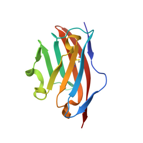
The crystal structure of myelin oligodendrocyte glycoprotein, a key autoantigen in multiple sclerosis

Clements, C.S.Reid, H.H.Beddoe, T.Tynan, F.E.Perugini, M.A.Johns, T.G.Bernard, C.C.Rossjohn, J.

(2003) Proc Natl Acad Sci U S A 100: 11059-11064

  • DOI: https://doi.org/10.1073/pnas.1833158100
  • Primary Citation Related Structures: 
    1PY9

  • PubMed Abstract: 

    Myelin oligodendrocyte glycoprotein (MOG) is a key CNS-specific autoantigen for primary demyelination in multiple sclerosis. Although the disease-inducing role of MOG has been established, its precise function in the CNS remains obscure. To gain new insights into the physiological and immunopathological role of MOG, we determined the 1.8-A crystal structure of the MOG extracellular domain (MOGED). MOGED adopts a classical Ig (Ig variable domain) fold that was observed to form an antiparallel head-to-tail dimer. A dimeric form of native MOG was observed, and MOGED was also shown to dimerize in solution, consistent with the view of MOG acting as a homophilic adhesion receptor. The MOG35-55 peptide, a major encephalitogenic determinant recognized by both T cells and demyelinating autoantibodies, is partly occluded within the dimer interface. The structure of this key autoantigen suggests a relationship between the dimeric form of MOG within the myelin sheath and a breakdown of immunological tolerance to MOG that is observed in multiple sclerosis.


  • Organizational Affiliation
    • Protein Crystallography Unit, Department of Biochemistry and Molecular Biology, School of Biomedical Sciences, Monash University, Clayton, Victoria 3168, Australia.

Macromolecule Content 

  • Total Structure Weight: 13.47 kDa 
  • Atom Count: 1,159 
  • Modeled Residue Count: 116 
  • Deposited Residue Count: 116 
  • Unique protein chains: 1

Macromolecules

Find similar proteins by:|  3D Structure
Entity ID: 1
MoleculeChains  Sequence LengthOrganismDetailsImage
Myelin-oligodendrocyte glycoprotein116Mus musculusMutation(s): 0 
Gene Names: MOG
UniProt
Find proteins for Q61885 (Mus musculus)
Explore Q61885 
Go to UniProtKB:  Q61885
Entity Groups
Sequence Clusters30% Identity50% Identity70% Identity90% Identity95% Identity100% Identity
UniProt GroupQ61885
Sequence Annotations
Expand
Reference Sequence

Experimental Data & Validation

Experimental Data

  • Method: X-RAY DIFFRACTION
  • Resolution: 1.80 Å
  • R-Value Free:  0.237 (Depositor), 0.236 (DCC) 
  • R-Value Work:  0.201 (Depositor), 0.202 (DCC) 
  • R-Value Observed: 0.201 (Depositor) 
Space Group: I 41
Unit Cell:
Length ( Å )Angle ( ˚ )
a = 65.72α = 90
b = 65.72β = 90
c = 67.58γ = 90
Software Package:
Software NamePurpose
AMoREphasing
CNSrefinement

Structure Validation

View Full Validation Report


Entry History 

Deposition Data

Revision History  (Full details and data files)

  • Version 1.0: 2003-09-30
    Type: Initial release
  • Version 1.1: 2008-04-29
    Changes: Version format compliance
  • Version 1.2: 2011-07-13
    Changes: Version format compliance
  • Version 1.3: 2017-10-11
    Changes: Refinement description
  • Version 1.4: 2019-12-11
    Changes: Data collection
  • Version 1.5: 2024-04-03
    Changes: Data collection, Database references, Derived calculations, Refinement description
  • Version 1.6: 2024-10-16
    Changes: Structure summary