1PUM | pdb_00001pum

Mistletoe lectin I in complex with galactose


Experimental Data Snapshot

  • Method: X-RAY DIFFRACTION
  • Resolution: 2.30 Å
  • R-Value Free: 
    0.246 (Depositor) 
  • R-Value Work: 
    0.209 (Depositor), 0.199 (DCC) 
  • R-Value Observed: 
    0.280 (Depositor) 

wwPDB Validation 3D Report Full Report

Validation slider image for 1PUM

Ligand Structure Quality Assessment 


This is version 2.1 of the entry. See complete history

Literature

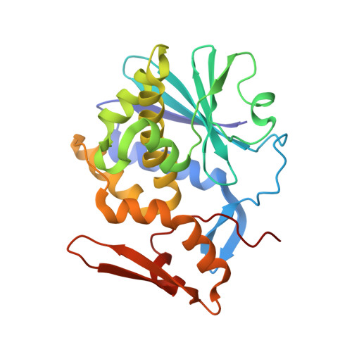
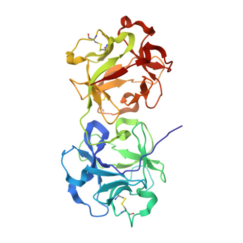
Mistletoe lectin I in complex with galactose and lactose reveals distinct sugar-binding properties

Mikeska, R.Wacker, R.Arni, R.Singh, T.P.Mikhailov, A.Gabdoulkhakov, A.Voelter, W.Betzel, C.

(2005) Acta Crystallogr Sect F Struct Biol Cryst Commun 61: 17-25

  • DOI: https://doi.org/10.1107/S1744309104031501
  • Primary Citation Related Structures: 
    1PUM, 1PUU

  • PubMed Abstract: 

    The structures of mistletoe lectin I (ML-I) from Viscum album complexed with lactose and galactose have been determined at 2.3 A resolution and refined to R factors of 20.9% (Rfree = 23.6%) and 20.9 (Rfree = 24.6%), respectively. ML-I is a heterodimer and belongs to the class of ribosome-inactivating proteins of type II, which consist of two chains. The A-chain has rRNA N-glycosidase activity and irreversibly inhibits eukaryotic ribosomes. The B-chain is a lectin and preferentially binds to galactose-terminated glycolipids and glycoproteins on cell membranes. Saccharide binding is performed by two binding sites in subdomains alpha1 and gamma2 of the ML-I B-chain separated by approximately 62 A from each other. The favoured binding of galactose in subdomain alpha1 is achieved via hydrogen bonds connecting the 4-hydroxyl and 3-hydroxyl groups of the sugar moiety with the side chains of Asp23B, Gln36B and Lys41B and the main chain of 26B. The aromatic ring of Trp38B on top of the preferred binding pocket supports van der Waals packing of the apolar face of galactose and stabilizes the sugar-lectin complex. In the galactose-binding site II of subdomain gamma2, Tyr249B provides the hydrophobic stacking and the side chains of Asp235B, Gln238B and Asn256B are hydrogen-bonding partners for galactose. In the case of the galactose-binding site I, the 2-hydroxyl group also stabilizes the sugar-protein complex, an interaction thus far rarely detected in galactose-specific lectins. Finally, a potential third low-affinity galactose-binding site in subunit beta1 was identified in the present ML-I structures, in which a glycerol molecule from the cryoprotectant buffer has bound, mimicking the sugar compound.


  • Organizational Affiliation
    • Institute of Biochemistry and Food Chemistry, University of Hamburg, c/o DESY, Notkestrasse 85, 22603 Hamburg, Germany.

Macromolecule Content 

  • Total Structure Weight: 59.25 kDa 
  • Atom Count: 4,505 
  • Modeled Residue Count: 512 
  • Deposited Residue Count: 512 
  • Unique protein chains: 2

Macromolecules

Find similar proteins by:|  3D Structure
Entity ID: 1
MoleculeChains  Sequence LengthOrganismDetailsImage
lectin I A chain249Viscum albumMutation(s): 0 
UniProt
Find proteins for P81446 (Viscum album)
Explore P81446 
Go to UniProtKB:  P81446
Entity Groups
Sequence Clusters30% Identity50% Identity70% Identity90% Identity95% Identity100% Identity
UniProt GroupP81446
Glycosylation
Glycosylation Sites: 1
Sequence Annotations
Expand
Reference Sequence
Find similar proteins by:|  3D Structure
Entity ID: 2
MoleculeChains  Sequence LengthOrganismDetailsImage
lectin I B chain263Viscum albumMutation(s): 0 
UniProt
Find proteins for P81446 (Viscum album)
Explore P81446 
Go to UniProtKB:  P81446
Entity Groups
Sequence Clusters30% Identity50% Identity70% Identity90% Identity95% Identity100% Identity
UniProt GroupP81446
Glycosylation
Glycosylation Sites: 3
Sequence Annotations
Expand
Reference Sequence

Oligosaccharides

Help  
Entity ID: 3
MoleculeChains Length2D Diagram GlycosylationD Interactions
2-acetamido-2-deoxy-alpha-D-glucopyranose-(1-4)-2-acetamido-2-deoxy-beta-D-glucopyranose
C
2N-Glycosylation
Glycosylation Resources
GlyTouCan: G07375KG
GlyCosmos: G07375KG
GlyGen: G07375KG
Entity ID: 4
MoleculeChains Length2D Diagram GlycosylationD Interactions
2-acetamido-2-deoxy-beta-D-glucopyranose-(1-4)-2-acetamido-2-deoxy-beta-D-glucopyranose
D
2N-Glycosylation
Glycosylation Resources
GlyTouCan: G42666HT
GlyCosmos: G42666HT
GlyGen: G42666HT
Entity ID: 5
MoleculeChains Length2D Diagram GlycosylationD Interactions
beta-L-fucopyranose-(1-3)-2-acetamido-2-deoxy-beta-D-glucopyranose
E
2N-Glycosylation
Glycosylation Resources
GlyTouCan: G95595RA
GlyCosmos: G95595RA
GlyGen: G95595RA

Small Molecules

Ligands 5 Unique
IDChains Name / Formula / InChI Key2D Diagram3D Interactions
NAG

Query on NAG


Download:Ideal Coordinates CCD File
F [auth A]2-acetamido-2-deoxy-beta-D-glucopyranose
C8 H15 N O6
OVRNDRQMDRJTHS-FMDGEEDCSA-N
GAL

Query on GAL


Download:Ideal Coordinates CCD File
R [auth B],
S [auth B]
beta-D-galactopyranose
C6 H12 O6
WQZGKKKJIJFFOK-FPRJBGLDSA-N
SO4

Query on SO4


Download:Ideal Coordinates CCD File
I [auth A],
J [auth A]
SULFATE ION
O4 S
QAOWNCQODCNURD-UHFFFAOYSA-L
GOL

Query on GOL


Download:Ideal Coordinates CCD File
AA [auth B]
BA [auth B]
K [auth A]
L [auth A]
M [auth A]
AA [auth B],
BA [auth B],
K [auth A],
L [auth A],
M [auth A],
N [auth A],
O [auth A],
P [auth A],
Q [auth A],
T [auth B],
U [auth B],
V [auth B],
W [auth B],
X [auth B],
Y [auth B],
Z [auth B]
GLYCEROL
C3 H8 O3
PEDCQBHIVMGVHV-UHFFFAOYSA-N
CL

Query on CL


Download:Ideal Coordinates CCD File
G [auth A],
H [auth A]
CHLORIDE ION
Cl
VEXZGXHMUGYJMC-UHFFFAOYSA-M

Experimental Data & Validation

Experimental Data

  • Method: X-RAY DIFFRACTION
  • Resolution: 2.30 Å
  • R-Value Free:  0.246 (Depositor) 
  • R-Value Work:  0.209 (Depositor), 0.199 (DCC) 
  • R-Value Observed: 0.280 (Depositor) 
Space Group: P 65 2 2
Unit Cell:
Length ( Å )Angle ( ˚ )
a = 106.826α = 90
b = 106.826β = 90
c = 310.951γ = 120
Software Package:
Software NamePurpose
DENZOdata reduction
SCALEPACKdata scaling
AMoREphasing
CNSrefinement

Structure Validation

View Full Validation Report


Ligand Structure Quality Assessment 


Entry History 

Deposition Data

Revision History  (Full details and data files)

  • Version 1.0: 2004-06-25
    Type: Initial release
  • Version 1.1: 2008-04-29
    Changes: Version format compliance
  • Version 1.2: 2011-07-13
    Changes: Non-polymer description, Version format compliance
  • Version 2.0: 2020-07-29
    Type: Remediation
    Reason: Carbohydrate remediation
    Changes: Atomic model, Data collection, Derived calculations, Structure summary
  • Version 2.1: 2024-10-09
    Changes: Data collection, Database references, Structure summary