1PSZ | pdb_00001psz

PNEUMOCOCCAL SURFACE ANTIGEN PSAA


Experimental Data Snapshot

  • Method: X-RAY DIFFRACTION
  • Resolution: 2.00 Å
  • R-Value Free: 
    0.236 (Depositor) 
  • R-Value Work: 
    0.178 (Depositor), 0.181 (DCC) 

wwPDB Validation 3D Report Full Report

Validation slider image for 1PSZ

This is version 1.4 of the entry. See complete history

Literature

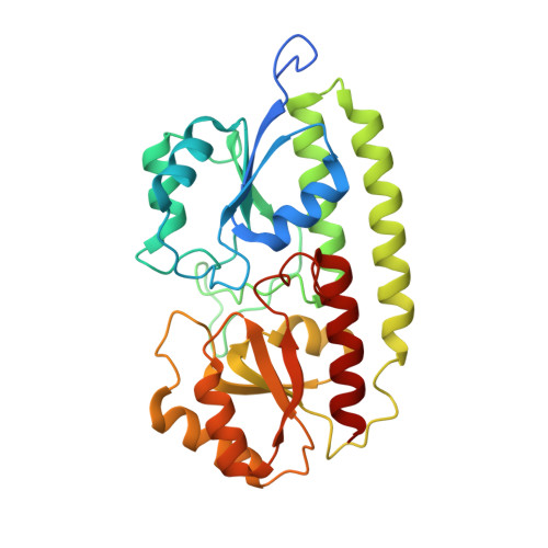
The crystal structure of pneumococcal surface antigen PsaA reveals a metal-binding site and a novel structure for a putative ABC-type binding protein.

Lawrence, M.C.Pilling, P.A.Epa, V.C.Berry, A.M.Ogunniyi, A.D.Paton, J.C.

(1998) Structure 6: 1553-1561

  • DOI: https://doi.org/10.1016/s0969-2126(98)00153-1
  • Primary Citation Related Structures: 
    1PSZ

  • PubMed Abstract: 

    . The surface protein PsaA of the pathogenic bacterium Streptococcus pneumoniae plays an essential role in its virulence. PsaA is a putative ATP-binding cassette-type (ABC-type) binding protein involved in the uptake of Mn2+ and possibly Zn2+ and is considered to be both a potential drug target and and a candidate vaccine component. . The structure of PsaA has been determined to 2.0 A resolution using X-ray crystallography and is the first structure obtained for an ABC-type binding protein from a Gram-positive organism. The protein consists of two (beta/alpha)4 domains linked together by a single helix. A metal-binding site is formed in the domain interface by the sidechains of His67, His139, Glu205 and Asp280 and is occupied in the structure. . The structural topology of PsaA is fundamentally different from that of other ABC-type binding proteins determined thus far in that PsaA lacks the characteristic 'hinge peptides' involved in conformational change upon solute uptake and release. In our structure, the metal-binding site is probably occupied by Zn2+. The site seems to be well conserved amongst related receptors from both Gram-positive and Gram-negative bacteria.


  • Organizational Affiliation
    • Biomolecular Research Institute 343 Royal Parade Parkville Victoria 3052 Australia. mike.lawrence@bioresi.com.au

Macromolecule Content 

  • Total Structure Weight: 34.21 kDa 
  • Atom Count: 2,502 
  • Modeled Residue Count: 286 
  • Deposited Residue Count: 303 
  • Unique protein chains: 1

Macromolecules

Find similar proteins by:|  3D Structure
Entity ID: 1
MoleculeChains  Sequence LengthOrganismDetailsImage
PROTEIN (SURFACE ANTIGEN PSAA)303Streptococcus pneumoniaeMutation(s): 0 
UniProt
Find proteins for P0A4G2 (Streptococcus pneumoniae serotype 4 (strain ATCC BAA-334 / TIGR4))
Explore P0A4G2 
Go to UniProtKB:  P0A4G2
Entity Groups
Sequence Clusters30% Identity50% Identity70% Identity90% Identity95% Identity100% Identity
UniProt GroupP0A4G2
Sequence Annotations
Expand
Reference Sequence

Small Molecules

Ligands 1 Unique
IDChains Name / Formula / InChI Key2D Diagram3D Interactions
ZN

Query on ZN


Download:Ideal Coordinates CCD File
B [auth A]ZINC ION
Zn
PTFCDOFLOPIGGS-UHFFFAOYSA-N

Experimental Data & Validation

Experimental Data

  • Method: X-RAY DIFFRACTION
  • Resolution: 2.00 Å
  • R-Value Free:  0.236 (Depositor) 
  • R-Value Work:  0.178 (Depositor), 0.181 (DCC) 
Space Group: P 21 21 21
Unit Cell:
Length ( Å )Angle ( ˚ )
a = 60.024α = 90
b = 66.517β = 90
c = 69.838γ = 90
Software Package:
Software NamePurpose
SHARPphasing
REFMACrefinement
DENZOdata reduction
SCALEPACKdata scaling

Structure Validation

View Full Validation Report


Entry History 

Deposition Data

Revision History  (Full details and data files)

  • Version 1.0: 2000-04-19
    Type: Initial release
  • Version 1.1: 2007-10-16
    Changes: Version format compliance
  • Version 1.2: 2011-07-13
    Changes: Version format compliance
  • Version 1.3: 2018-04-04
    Changes: Data collection
  • Version 1.4: 2023-12-27
    Changes: Data collection, Database references, Derived calculations