1PRE | pdb_00001pre

PROAEROLYSIN


Experimental Data Snapshot

  • Method: X-RAY DIFFRACTION
  • Resolution: 2.80 Å

wwPDB Validation 3D Report Full Report

Validation slider image for 1PRE

This is version 1.3 of the entry. See complete history

Literature

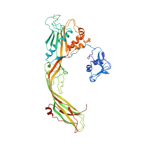
Structure of the Aeromonas toxin proaerolysin in its water-soluble and membrane-channel states.

Parker, M.W.Buckley, J.T.Postma, J.P.Tucker, A.D.Leonard, K.Pattus, F.Tsernoglou, D.

(1994) Nature 367: 292-295

  • DOI: https://doi.org/10.1038/367292a0
  • Primary Citation Related Structures: 
    1PRE

  • PubMed Abstract: 

    Aerolysin is chiefly responsible for the pathogenicity of Aeromonas hydrophila, a bacterium associated with diarrhoeal diseases and deep wound infections. Like many other microbial toxins, the protein changes in a multistep process from a completely water-soluble form to produce a transmembrane channel that destroys sensitive cells by breaking their permeability barriers. Here we describe the structure of proaerolysin determined by X-ray crystallography at 2.8 A resolution. The protoxin (M(r) 52,000) adopts a novel protein fold. Images of an aerolysin oligomer derived from electron microscopy have assisted in constructing a model of the membrane channel and have led to the proposal of a scheme to account for insertion of the protein into lipid bilayers to form ion channels.


  • Organizational Affiliation
    • St Vincent's Institute of Medical Research, Fitzroy, Victoria, Australia.

Macromolecule Content 

  • Total Structure Weight: 103.96 kDa 
  • Atom Count: 7,089 
  • Modeled Residue Count: 900 
  • Deposited Residue Count: 940 
  • Unique protein chains: 1

Macromolecules

Find similar proteins by:|  3D Structure
Entity ID: 1
MoleculeChains  Sequence LengthOrganismDetailsImage
PROAEROLYSIN
A, B
470Aeromonas hydrophilaMutation(s): 0 
UniProt
Find proteins for P09167 (Aeromonas hydrophila)
Explore P09167 
Go to UniProtKB:  P09167
Entity Groups
Sequence Clusters30% Identity50% Identity70% Identity90% Identity95% Identity100% Identity
UniProt GroupP09167
Sequence Annotations
Expand
Reference Sequence

Experimental Data & Validation

Experimental Data

  • Method: X-RAY DIFFRACTION
  • Resolution: 2.80 Å
Space Group: P 43 21 2
Unit Cell:
Length ( Å )Angle ( ˚ )
a = 104α = 90
b = 104β = 90
c = 222γ = 90
Software Package:
Software NamePurpose
MOSFLMdata reduction
PROLSQrefinement

Structure Validation

View Full Validation Report


Entry History 

Deposition Data

Revision History  (Full details and data files)

  • Version 1.0: 1996-10-14
    Type: Initial release
  • Version 1.1: 2008-03-24
    Changes: Version format compliance
  • Version 1.2: 2011-07-13
    Changes: Version format compliance
  • Version 1.3: 2024-10-16
    Changes: Data collection, Database references, Structure summary