1PPG | pdb_00001ppg

The refined 2.3 angstroms crystal structure of human leukocyte elastase in a complex with a valine chloromethyl ketone inhibitor


Experimental Data Snapshot

  • Method: X-RAY DIFFRACTION
  • Resolution: 2.30 Å
  • R-Value Work: 
    0.145 (Depositor) 

wwPDB Validation 3D Report Full Report

Validation slider image for 1PPG

This is version 3.0 of the entry. See complete history

Literature

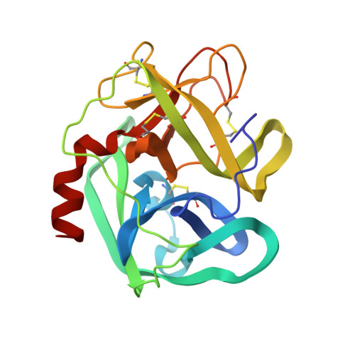
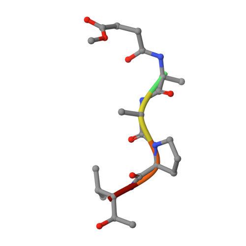
The refined 2.3 A crystal structure of human leukocyte elastase in a complex with a valine chloromethyl ketone inhibitor.

Wei, A.Z.Mayr, I.Bode, W.

(1988) FEBS Lett 234: 367-373

  • DOI: https://doi.org/10.1016/0014-5793(88)80118-2
  • Primary Citation Related Structures: 
    1PPG

  • PubMed Abstract: 

    The stoichiometric complex formed between human leukocyte elastase and a synthetic MeO-Suc-Ala-Ala-Pro-Val chloromethyl ketone inhibitor was co-crystallized and its X-ray structure determined, using Patterson search methods. Its structure has been crystallographically refined to a final R value of 0.145 (8.0 and 2.3 A). The enzyme structure is very similar to that recently observed in a complex formed with the ovomucoid third domain from turkey [(1986) EMBO J. 5,2453-2458]. The rms deviation of all alpha-carbon atoms is 0.32 A. The peptidic inhibitor is bound in a similar overall conformation as the ovomucoid binding segment. Covalent bonds are formed between Val-P1 of the inhibitor and His-57 NE2 and Ser-195 OG of the enzyme. The carbonyl carbon is tetrahedrally deformed to a hemiketal. The valine side chain is arranged in the S1 pocket in the g-conformation.


  • Organizational Affiliation
    • Max-Planck-Institut für Biochemie, Martinsried, FRG.

Macromolecule Content 

  • Total Structure Weight: 26.67 kDa 
  • Atom Count: 2,053 
  • Modeled Residue Count: 224 
  • Deposited Residue Count: 224 
  • Unique protein chains: 2

Macromolecules

Find similar proteins by:|  3D Structure
Entity ID: 1
MoleculeChains  Sequence LengthOrganismDetailsImage
HUMAN LEUKOCYTE ELASTASEA [auth E]218Homo sapiensMutation(s): 0 
EC: 3.4.21.37
UniProt & NIH Common Fund Data Resources
Find proteins for P08246 (Homo sapiens)
Explore P08246 
Go to UniProtKB:  P08246
PHAROS:  P08246
GTEx:  ENSG00000197561 
Entity Groups
Sequence Clusters30% Identity50% Identity70% Identity90% Identity95% Identity100% Identity
UniProt GroupP08246
Glycosylation
Glycosylation Sites: 2Go to GlyGen: P08246-1
Sequence Annotations
Expand
Reference Sequence
Find similar proteins by:  Sequence   |   3D Structure  
Entity ID: 2
MoleculeChains  Sequence LengthOrganismDetailsImage
MEO-SUCCINYL-ALA-ALA-PRO-VAL CHLOROMETHYLKETONEB [auth I]6N/AMutation(s): 0 

Oligosaccharides

Help  
Entity ID: 3
MoleculeChains Length2D Diagram GlycosylationD Interactions
beta-D-galactopyranose-(1-4)-2-acetamido-2-deoxy-beta-D-glucopyranose-(1-2)-alpha-D-mannopyranose-(1-6)-[alpha-D-mannopyranose-(1-3)]beta-D-mannopyranose-(1-4)-2-acetamido-2-deoxy-beta-D-glucopyranose-(1-4)-[alpha-L-fucopyranose-(1-6)]2-acetamido-2-deoxy-beta-D-glucopyranoseC [auth A]8N-Glycosylation
Glycosylation Resources
GlyTouCan: G61334IA
GlyCosmos: G61334IA
GlyGen: G61334IA
Entity ID: 4
MoleculeChains Length2D Diagram GlycosylationD Interactions
alpha-D-glucopyranose-(1-4)-2-acetamido-2-deoxy-alpha-D-glucopyranose-(1-2)-beta-D-mannopyranose-(1-6)-[alpha-D-mannopyranose-(1-3)]beta-D-mannopyranose-(1-4)-2-acetamido-2-deoxy-beta-D-glucopyranose-(1-4)-[alpha-L-fucopyranose-(1-6)]2-acetamido-2-deoxy-beta-D-glucopyranoseD [auth B]8N-Glycosylation
Glycosylation Resources
GlyTouCan: G32078TL
GlyCosmos: G32078TL
GlyGen: G32078TL

Small Molecules

Modified Residues  1 Unique
IDChains TypeFormula2D DiagramParent
VAI
Query on VAI
B [auth I]PEPTIDE-LIKEC5 H13 N O2VAL

Biologically Interesting Molecules (External Reference) 

1 Unique

Experimental Data & Validation

Experimental Data

  • Method: X-RAY DIFFRACTION
  • Resolution: 2.30 Å
  • R-Value Work:  0.145 (Depositor) 
Space Group: P 63
Unit Cell:
Length ( Å )Angle ( ˚ )
a = 74.2α = 90
b = 74.2β = 90
c = 70.6γ = 120
Software Package:
Software NamePurpose
EREFrefinement

Structure Validation

View Full Validation Report


Entry History 

Deposition Data

Revision History  (Full details and data files)

  • Version 1.0: 1994-01-31
    Type: Initial release
  • Version 1.1: 2008-03-24
    Changes: Version format compliance
  • Version 1.2: 2011-07-13
    Changes: Atomic model, Database references, Derived calculations, Non-polymer description, Structure summary, Version format compliance
  • Version 1.3: 2012-12-12
    Changes: Other
  • Version 2.0: 2020-07-29
    Type: Remediation
    Reason: Carbohydrate remediation
    Changes: Advisory, Atomic model, Data collection, Derived calculations, Other, Structure summary
  • Version 3.0: 2024-07-10
    Changes: Advisory, Data collection, Database references, Polymer sequence, Structure summary