1POC | pdb_00001poc

CRYSTAL STRUCTURE OF BEE-VENOM PHOSPHOLIPASE A2 IN A COMPLEX WITH A TRANSITION-STATE ANALOGUE


Experimental Data Snapshot

  • Method: X-RAY DIFFRACTION
  • Resolution: 2.00 Å
  • R-Value Work: 
    0.192 (Depositor) 
  • R-Value Observed: 
    0.192 (Depositor) 

wwPDB Validation 3D Report Full Report

Validation slider image for 1POC

This is version 1.5 of the entry. See complete history

Literature

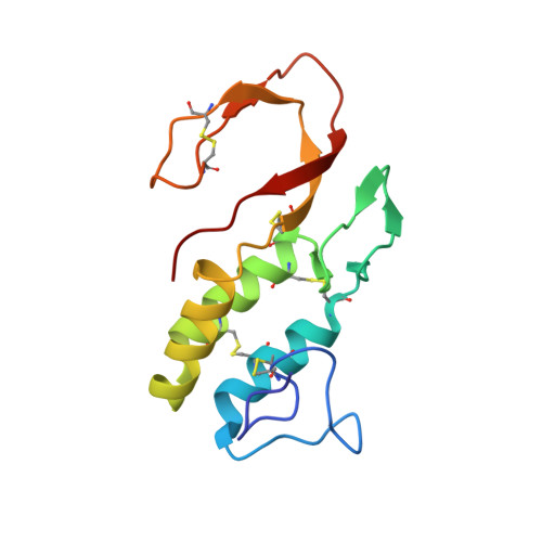
Crystal structure of bee-venom phospholipase A2 in a complex with a transition-state analogue.

Scott, D.L.Otwinowski, Z.Gelb, M.H.Sigler, P.B.

(1990) Science 250: 1563-1566

  • DOI: https://doi.org/10.1126/science.2274788
  • Primary Citation Related Structures: 
    1POC

  • PubMed Abstract: 

    The 2.0 angstroms crystal structure of a complex containing bee-venom phospholipase A2 (PLA2) and a phosphonate transition-state analogue was solved by multiple isomorphous replacement. The electron-density map is sufficiently detailed to visualize the proximal sugars of the enzyme's N-linked carbohydrate and a single molecule of the transition-state analogue bound ot its active center. Although bee-venom PLA2 does not belong to the large homologous Class I/II family that encompasses most other well-studied PLA2s, there is segmental sequence similarity and conservation of many functional substructures. Comparison of the bee-venom enzyme with other phospholipase structures provides compelling evidence for a common catalytic mechanism.


  • Organizational Affiliation
    • Department of Molecular Biophysics and Biochemistry, Yale University, New Haven, CT 06511.

Macromolecule Content 

  • Total Structure Weight: 15.8 kDa 
  • Atom Count: 1,177 
  • Modeled Residue Count: 134 
  • Deposited Residue Count: 134 
  • Unique protein chains: 1

Macromolecules

Find similar proteins by:|  3D Structure
Entity ID: 1
MoleculeChains  Sequence LengthOrganismDetailsImage
PHOSPHOLIPASE A2134Apis melliferaMutation(s): 0 
EC: 3.1.1.4
Membrane Entity: Yes 
UniProt
Find proteins for P00630 (Apis mellifera)
Explore P00630 
Go to UniProtKB:  P00630
Entity Groups
Sequence Clusters30% Identity50% Identity70% Identity90% Identity95% Identity100% Identity
UniProt GroupP00630
Sequence Annotations
Expand
Reference Sequence

Small Molecules

Ligands 2 Unique
IDChains Name / Formula / InChI Key2D Diagram3D Interactions
GEL

Query on GEL


Download:Ideal Coordinates CCD File
C [auth A]1-O-OCTYL-2-HEPTYLPHOSPHONYL-SN-GLYCERO-3-PHOSPHOETHANOLAMINE
C20 H45 N O8 P2
RCCNUBYROFOKAU-HXUWFJFHSA-N
CA

Query on CA


Download:Ideal Coordinates CCD File
B [auth A]CALCIUM ION
Ca
BHPQYMZQTOCNFJ-UHFFFAOYSA-N

Experimental Data & Validation

Experimental Data

  • Method: X-RAY DIFFRACTION
  • Resolution: 2.00 Å
  • R-Value Work:  0.192 (Depositor) 
  • R-Value Observed: 0.192 (Depositor) 
Space Group: I 41 2 2
Unit Cell:
Length ( Å )Angle ( ˚ )
a = 89.5α = 90
b = 89.5β = 90
c = 132.5γ = 90
Software Package:
Software NamePurpose
X-PLORmodel building
PROFFTrefinement
X-PLORrefinement
X-PLORphasing

Structure Validation

View Full Validation Report


Entry History 

Deposition Data

Revision History  (Full details and data files)

  • Version 1.0: 1993-10-31
    Type: Initial release
  • Version 1.1: 2008-03-03
    Changes: Version format compliance
  • Version 1.2: 2011-07-13
    Changes: Version format compliance
  • Version 1.3: 2019-07-17
    Changes: Data collection, Other, Refinement description
  • Version 1.4: 2019-08-14
    Changes: Data collection, Refinement description
  • Version 1.5: 2024-10-30
    Changes: Data collection, Database references, Derived calculations, Structure summary