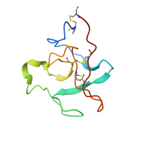
The structure of recombinant plasminogen kringle 1 and the fibrin binding site.
Wu, T.P., Padmanabhan, K.P., Tulinsky, A.(1994) Blood Coagul Fibrinolysis 5: 157-166
- PubMed: 8054447
- DOI: https://doi.org/10.1097/00001721-199404000-00001
- Primary Citation of Related Structures:
1PKR - PubMed Abstract:
The structure of recombinant (Hoover et al. Biochemistry, 1993; 32:10936-10944) plasminogen (PG) kringle 1 (K1) has been determined and refined at 2.48 A resolution to a crystallographic R value of 0.159. In addition, 71 water molecules and two chloride ions have been located. The folding of PGK1 is very similar to that of PGK4. The lysine/fibrin binding site, however, differs from that of both PGK4 and tissue-type PG activator (t-PA) K2 at the cationic centre. Although PGK1 can potentially have a doubly charged cationic centre utilizing Arg34 and Arg71, the side chain of Arg34 is outside of Arg71 in a solvent region and its guanidino group is flexibly disordered. Moreover, site specific mutagenesis studies show unequivocally that Arg34 can be changed to glutamine without affecting the binding ability of PGK1. Thus, PGK1 only has Arg71 at the cationic site, PGK4 has Lys35/Arg71 and t-PAK2 has only Lys33. The cationic site differences may result in subtle responses in the binding affinities of the kringles. The two chloride ions are located in the lysine binding site and effectively compensate the positive charges of the region. They also appear to be involved intermolecularly in a complex way in the crystal structure. Such intermolecular anionic interactions are also found in PGK4 and t-PAK2.
Organizational Affiliation:
Department of Chemistry, Michigan State University, East Lansing 48824.


