1PKM | pdb_00001pkm

THE REFINED THREE-DIMENSIONAL STRUCTURE OF CAT MUSCLE (M1) PYRUVATE KINASE, AT A RESOLUTION OF 2.6 ANGSTROMS


Experimental Data Snapshot

  • Method: X-RAY DIFFRACTION
  • Resolution: 2.60 Å
  • R-Value Work: 
    0.175 (Depositor) 

wwPDB Validation 3D Report Full Report

Validation slider image for 1PKM

This is version 1.5 of the entry. See complete history

Literature

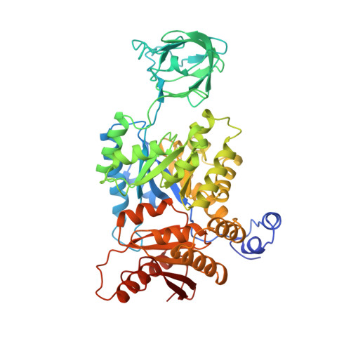
Refined three-dimensional structure of cat-muscle (M1) pyruvate kinase at a resolution of 2.6 A.

Allen, S.C.Muirhead, H.

(1996) Acta Crystallogr D Biol Crystallogr 52: 499-504

  • DOI: https://doi.org/10.1107/S0907444995016040
  • Primary Citation Related Structures: 
    1PKM

  • PubMed Abstract: 

    The three-dimensional structure of cat-muscle pyoruvate kinase has been refined at a resolution of 2.6 A. The details of the structure permit interpretation of the original heavy-atom studies and give insight into the importance of conserved residues in pyruvate kinases and the allosteric behaviour of the enzyme. There are a small number of essential residues which determine the relative orientations of domains and the precise nature of intersubunit contacts. Arginine residues are particularly important.


  • Organizational Affiliation
    • Department of Biochemistry and Molecular Recognition Centre, School of Medical Sciences, University of Bristol.

Macromolecule Content 

  • Total Structure Weight: 58 kDa 
  • Atom Count: 3,976 
  • Modeled Residue Count: 519 
  • Deposited Residue Count: 530 
  • Unique protein chains: 1

Macromolecules

Find similar proteins by:|  3D Structure
Entity ID: 1
MoleculeChains  Sequence LengthOrganismDetailsImage
M1 PYRUVATE KINASE530Felis catusMutation(s): 0 
EC: 2.7.1.40 (PDB Primary Data), 2.7.11.1 (UniProt), 2.7.10.2 (UniProt)
UniProt
Find proteins for P11979 (Felis catus)
Explore P11979 
Go to UniProtKB:  P11979
Entity Groups
Sequence Clusters30% Identity50% Identity70% Identity90% Identity95% Identity100% Identity
UniProt GroupP11979
Sequence Annotations
Expand
Reference Sequence

Experimental Data & Validation

Experimental Data

  • Method: X-RAY DIFFRACTION
  • Resolution: 2.60 Å
  • R-Value Work:  0.175 (Depositor) 
Space Group: I 2 2 2
Unit Cell:
Length ( Å )Angle ( ˚ )
a = 88.4α = 90
b = 115.3β = 90
c = 131γ = 90
Software Package:
Software NamePurpose
X-PLORmodel building
TNTrefinement
X-PLORrefinement
MOSCOdata reduction
3DSCALEdata reduction
X-PLORphasing

Structure Validation

View Full Validation Report


Entry History 

Deposition Data

Revision History  (Full details and data files)

  • Version 1.0: 1995-10-15
    Type: Initial release
  • Version 1.1: 2008-03-24
    Changes: Version format compliance
  • Version 1.2: 2011-07-13
    Changes: Derived calculations, Version format compliance
  • Version 1.3: 2019-07-17
    Changes: Data collection, Other, Refinement description
  • Version 1.4: 2019-08-14
    Changes: Data collection, Refinement description
  • Version 1.5: 2024-02-14
    Changes: Data collection, Database references