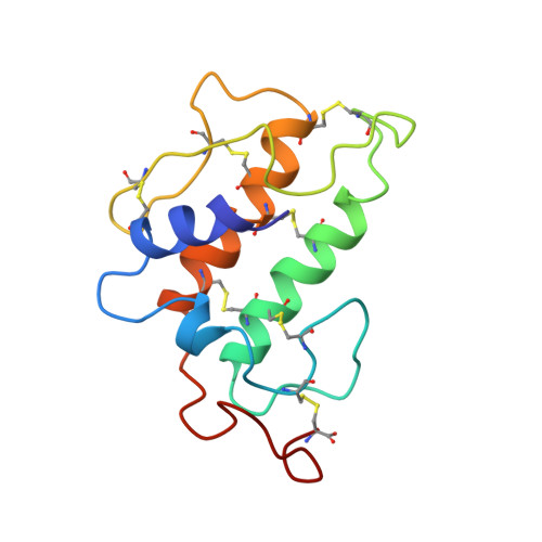
Solution structure of porcine pancreatic phospholipase A2.
van den Berg, B., Tessari, M., de Haas, G.H., Verheij, H.M., Boelens, R., Kaptein, R.(1995) EMBO J 14: 4123-4131
- PubMed: 7556053 Search on PubMedSearch on PubMed Central
- DOI: https://doi.org/10.1002/j.1460-2075.1995.tb00086.x
- Primary Citation Related Structures:
1PIR, 1PIS - PubMed Abstract:
The lipolytic enzyme phospholipase A2 (PLA2) is involved in the degradation of high-molecular weight phospholipid aggregates in vivo. The enzyme has very high catalytic activities on aggregated substrates compared with monomeric substrates, a phenomenon called interfacial activation. Crystal structures of PLA2s in the absence and presence of inhibitors are identical, from which it has been concluded that enzymatic conformational changes do not play a role in the mechanism of interfacial activation. The high-resolution NMR structure of porcine pancreatic PLA2 free in solution was determined with heteronuclear multidimensional NMR methodology using doubly labeled 13C, 15N-labeled protein. The solution structure of PLA2 shows important deviations from the crystal structure. In the NMR structure the Ala1 alpha-amino group is disordered and the hydrogen bonding network involving the N-terminus and the active site is incomplete. The disorder observed for the N-terminal region of PLA2 in the solution structure could be related to the low activity of the enzyme towards monomeric substrates. The NMR structure of PLA2 suggests, in contrast to the crystallographic work, that conformational changes do play a role in the interfacial activation of this enzyme.
- Center for Biomembranes and Lipid Enzymology, Utrecht University, The Netherlands.
Organizational Affiliation: