1PHN | pdb_00001phn

STRUCTURE OF PHYCOCYANIN FROM CYANIDIUM CALDARIUM AT 1.65A RESOLUTION


Experimental Data Snapshot

  • Method: X-RAY DIFFRACTION
  • Resolution: 1.65 Å
  • R-Value Free: 
    0.272 (Depositor) 
  • R-Value Work: 
    0.183 (Depositor), 0.189 (DCC) 
  • R-Value Observed: 
    0.183 (Depositor) 

wwPDB Validation 3D Report Full Report

Validation slider image for 1PHN

Ligand Structure Quality Assessment 


This is version 1.3 of the entry. See complete history

Literature

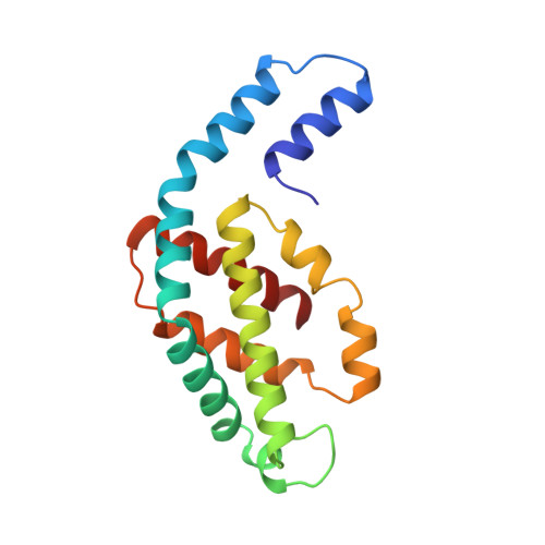
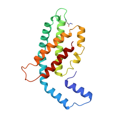
Crystal structure of C-phycocyanin from Cyanidium caldarium provides a new perspective on phycobilisome assembly.

Stec, B.Troxler, R.F.Teeter, M.M.

(1999) Biophys J 76: 2912-2921

  • DOI: https://doi.org/10.1016/S0006-3495(99)77446-1
  • Primary Citation Related Structures: 
    1PHN

  • PubMed Abstract: 

    The crystal structure of the light-harvesting protein phycocyanin from the cyanobacterium Cyanidium caldarium with novel crystal packing has been solved at 1.65-A resolution. The structure has been refined to an R value of 18.3% with excellent backbone and side-chain stereochemical parameters. In crystals of phycocyanin used in this study, the hexamers are offset rather than aligned as in other phycocyanins that have been crystallized to date. Analysis of this crystal's unique packing leads to a proposal for phycobilisome assembly in vivo and for a more prominent role for chromophore beta-155. This new role assigned to chromophore beta-155 in phycocyanin sheds light on the numerical relationships among and function of external chromophores found in phycoerythrins and phycoerythrocyanins.


  • Organizational Affiliation
    • Department of Chemistry, Merkert Chemistry Building, Boston College, Chestnut Hill, Massachusetts 02167, USA. stec@bioc.rice.edu

Macromolecule Content 

  • Total Structure Weight: 37.57 kDa 
  • Atom Count: 3,208 
  • Modeled Residue Count: 334 
  • Deposited Residue Count: 334 
  • Unique protein chains: 2

Macromolecules

Find similar proteins by:|  3D Structure
Entity ID: 1
MoleculeChains  Sequence LengthOrganismDetailsImage
PHYCOCYANIN162Cyanidium caldariumMutation(s): 0 
UniProt
Find proteins for P00306 (Galdieria sulphuraria)
Explore P00306 
Go to UniProtKB:  P00306
Entity Groups
Sequence Clusters30% Identity50% Identity70% Identity90% Identity95% Identity100% Identity
UniProt GroupP00306
Sequence Annotations
Expand
Reference Sequence
Find similar proteins by:|  3D Structure
Entity ID: 2
MoleculeChains  Sequence LengthOrganismDetailsImage
PHYCOCYANIN172Cyanidium caldariumMutation(s): 0 
UniProt
Find proteins for P00311 (Galdieria sulphuraria)
Explore P00311 
Go to UniProtKB:  P00311
Entity Groups
Sequence Clusters30% Identity50% Identity70% Identity90% Identity95% Identity100% Identity
UniProt GroupP00311
Sequence Annotations
Expand
Reference Sequence

Small Molecules

Experimental Data & Validation

Experimental Data

  • Method: X-RAY DIFFRACTION
  • Resolution: 1.65 Å
  • R-Value Free:  0.272 (Depositor) 
  • R-Value Work:  0.183 (Depositor), 0.189 (DCC) 
  • R-Value Observed: 0.183 (Depositor) 
Space Group: H 3 2
Unit Cell:
Length ( Å )Angle ( ˚ )
a = 106.42α = 90
b = 106.42β = 90
c = 176.18γ = 120
Software Package:
Software NamePurpose
X-PLORmodel building
X-PLORrefinement
SDMSdata reduction
X-PLORphasing

Structure Validation

View Full Validation Report


Ligand Structure Quality Assessment 


Entry History 

Deposition Data

Revision History  (Full details and data files)

  • Version 1.0: 1997-09-17
    Type: Initial release
  • Version 1.1: 2008-03-25
    Changes: Version format compliance
  • Version 1.2: 2011-07-13
    Changes: Version format compliance
  • Version 1.3: 2024-06-05
    Changes: Data collection, Database references, Derived calculations, Other