1PHC | pdb_00001phc

CRYSTAL STRUCTURE OF SUBSTRATE-FREE PSEUDOMONAS PUTIDA CYTOCHROME P450


Experimental Data Snapshot

  • Method: X-RAY DIFFRACTION
  • Resolution: 1.60 Å
  • R-Value Observed: 
    0.190 (Depositor) 

wwPDB Validation 3D Report Full Report

Validation slider image for 1PHC

This is version 1.4 of the entry. See complete history

Literature

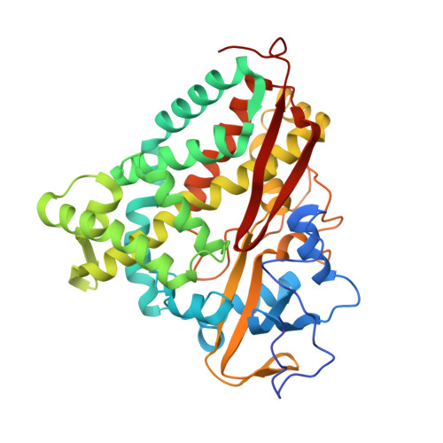
Crystal structure of substrate-free Pseudomonas putida cytochrome P-450.

Poulos, T.L.Finzel, B.C.Howard, A.J.

(1986) Biochemistry 25: 5314-5322

  • DOI: https://doi.org/10.1021/bi00366a049
  • Primary Citation Related Structures: 
    1PHC

  • PubMed Abstract: 

    The crystal structure of Pseudomonas putida cytochrome P-450cam in the substrate-free form has been refined at 2.20-A resolution and compared to the substrate-bound form of the enzyme. In the absence of the substrate camphor, the P-450cam heme iron atom is hexacoordinate with the sulfur atom of Cys-357 providing one axial heme ligand and a water molecule or hydroxide ion providing the other axial ligand. A network of hydrogen-bonded solvent molecules occupies the substrate pocket in addition to the iron-linked aqua ligand. When a camphor molecule binds, the active site waters including the aqua ligand are displaced, resulting in a pentacoordinate high-spin heme iron atom. Analysis of the Fno camphor - F camphor difference Fourier and a quantitative comparison of the two refined structures reveal that no detectable conformational change results from camphor binding other than a small repositioning of a phenylalanine side chain that contacts the camphor molecule. However, large decreases in the mean temperature factors of three separate segments of the protein centered on Tyr-96, Thr-185, and Asp-251 result from camphor binding. This indicates that camphor binding decreases the flexibility in these three regions of the P-450cam molecule without altering the mean position of the atoms involved.

Macromolecule Content 

  • Total Structure Weight: 47.21 kDa 
  • Atom Count: 3,439 
  • Modeled Residue Count: 405 
  • Deposited Residue Count: 414 
  • Unique protein chains: 1

Macromolecules

Find similar proteins by:|  3D Structure
Entity ID: 1
MoleculeChains  Sequence LengthOrganismDetailsImage
CYTOCHROME P450-CAM414Pseudomonas putidaMutation(s): 0 
EC: 1.14.15.1
UniProt
Find proteins for P00183 (Pseudomonas putida)
Explore P00183 
Go to UniProtKB:  P00183
Entity Groups
Sequence Clusters30% Identity50% Identity70% Identity90% Identity95% Identity100% Identity
UniProt GroupP00183
Sequence Annotations
Expand
Reference Sequence

Small Molecules

Ligands 1 Unique
IDChains Name / Formula / InChI Key2D Diagram3D Interactions
HEM

Query on HEM


Download:Ideal Coordinates CCD File
B [auth A]PROTOPORPHYRIN IX CONTAINING FE
C34 H32 Fe N4 O4
KABFMIBPWCXCRK-RGGAHWMASA-L

Experimental Data & Validation

Experimental Data

  • Method: X-RAY DIFFRACTION
  • Resolution: 1.60 Å
  • R-Value Observed: 0.190 (Depositor) 
Space Group: P 21 21 21
Unit Cell:
Length ( Å )Angle ( ˚ )
a = 108.67α = 90
b = 103.9β = 90
c = 36.38γ = 90
Software Package:
Software NamePurpose
PROLSQrefinement

Structure Validation

View Full Validation Report


Entry History 

Deposition Data

  • Released Date: 1993-10-31 
  • Deposition Author(s): Poulos, T.L.

Revision History  (Full details and data files)

  • Version 1.0: 1993-10-31
    Type: Initial release
  • Version 1.1: 2008-03-03
    Changes: Version format compliance
  • Version 1.2: 2011-07-13
    Changes: Version format compliance
  • Version 1.3: 2017-11-29
    Changes: Derived calculations, Other
  • Version 1.4: 2024-02-14
    Changes: Data collection, Database references, Derived calculations