1PHA | pdb_00001pha

INHIBITOR-INDUCED CONFORMATIONAL CHANGE IN CYTOCHROME P450-CAM


Experimental Data Snapshot

  • Method: X-RAY DIFFRACTION
  • Resolution: 1.63 Å
  • R-Value Observed: 
    0.190 (Depositor) 

wwPDB Validation 3D Report Full Report

Validation slider image for 1PHA

This is version 1.4 of the entry. See complete history

Literature

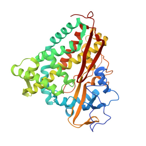
Inhibitor-induced conformational change in cytochrome P-450CAM.

Raag, R.Li, H.Jones, B.C.Poulos, T.L.

(1993) Biochemistry 32: 4571-4578

  • DOI: https://doi.org/10.1021/bi00068a013
  • Primary Citation Related Structures: 
    1PHA, 1PHB

  • PubMed Abstract: 

    The X-ray crystal structures of cytochrome P-450CAM complexed with both enantiomers of a chiral, multifunctional inhibitor have been refined to R-factors of 21.0% [(+)-enantiomer] and 19.6% [(-)-enantiomer] at approximately 2.1-A resolution. Binding of either enantiomer, both considerably larger than the natural substrate camphor, results in similar, dramatic structural changes in the enzyme. In contrast to all previous P-450CAM crystallographic structures, the Tyr96 side chain is not pointing "down" toward the heme but is rather directed "up" into the proposed substrate access channel. This conformational change is accompanied by the displacement of the Phe193 side chain out into the solvent at the enzyme surface. These changes are consistent with the assignment of this region of the enzyme as the access channel [Poulos et al. (1986) Biochemistry 25, 5314-5322] and suggest that several aromatic residues lining the channel may be involved in substrate recognition and channeling to the active site. The cation usually observed coordinated to the Tyr96 carbonyl oxygen is missing in the presence of the (+)-enantiomer but is present with the (-)-enantiomer. The Phe87 side chain, located near the inhibitor binding site, adopts different orientations depending upon which enantiomer is bound. Finally, electron density reveals that although the inhibitor enantiomers were dichlorinated as provided, when bound to P-450CAM the chlorine atoms are present at only 0-20% occupancy, probably reflecting selective binding of impurities in the samples. Coordinates of these inhibited P-450CAM complexes have been deposited in the Brookhaven Protein Data Bank [Bernstein et al. (1977) J. Mol. Biol. 112, 535-542].


  • Organizational Affiliation
    • Center for Advanced Research in Biotechnology, Maryland Biotechnology Institute, University of Maryland, Rockville 20850.

Macromolecule Content 

  • Total Structure Weight: 47.55 kDa 
  • Atom Count: 3,482 
  • Modeled Residue Count: 405 
  • Deposited Residue Count: 414 
  • Unique protein chains: 1

Macromolecules

Find similar proteins by:|  3D Structure
Entity ID: 1
MoleculeChains  Sequence LengthOrganismDetailsImage
CYTOCHROME P450-CAM414Pseudomonas putidaMutation(s): 0 
EC: 1.14.15.1
UniProt
Find proteins for P00183 (Pseudomonas putida)
Explore P00183 
Go to UniProtKB:  P00183
Entity Groups
Sequence Clusters30% Identity50% Identity70% Identity90% Identity95% Identity100% Identity
UniProt GroupP00183
Sequence Annotations
Expand
Reference Sequence

Small Molecules

Ligands 2 Unique
IDChains Name / Formula / InChI Key2D Diagram3D Interactions
HEM

Query on HEM


Download:Ideal Coordinates CCD File
B [auth A]PROTOPORPHYRIN IX CONTAINING FE
C34 H32 Fe N4 O4
KABFMIBPWCXCRK-RGGAHWMASA-L
PFZ

Query on PFZ


Download:Ideal Coordinates CCD File
C [auth A]1-(N-IMIDAZOLYL)-2-HYDROXY-2-(2,3-DICHLOROPHENYL)OCTANE
C17 H22 Cl2 N2 O
WJBNLGBHMWNHRZ-KRWDZBQOSA-N

Experimental Data & Validation

Experimental Data

  • Method: X-RAY DIFFRACTION
  • Resolution: 1.63 Å
  • R-Value Observed: 0.190 (Depositor) 
Space Group: P 21 21 21
Unit Cell:
Length ( Å )Angle ( ˚ )
a = 108.67α = 90
b = 103.9β = 90
c = 36.38γ = 90
Software Package:
Software NamePurpose
PROLSQrefinement

Structure Validation

View Full Validation Report


Entry History 

Deposition Data

  • Released Date: 1993-10-31 
  • Deposition Author(s): Poulos, T.L.

Revision History  (Full details and data files)

  • Version 1.0: 1993-10-31
    Type: Initial release
  • Version 1.1: 2008-03-03
    Changes: Version format compliance
  • Version 1.2: 2011-07-13
    Changes: Version format compliance
  • Version 1.3: 2017-11-29
    Changes: Derived calculations, Other
  • Version 1.4: 2024-02-14
    Changes: Data collection, Database references, Derived calculations